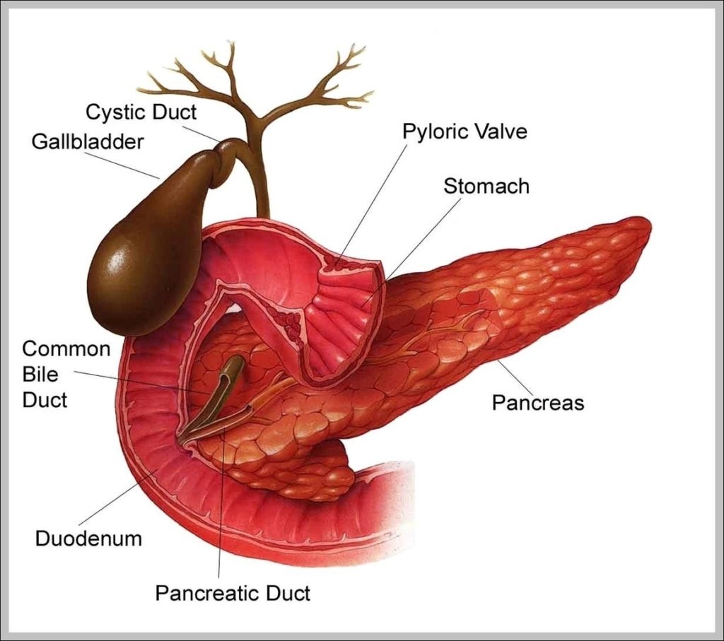
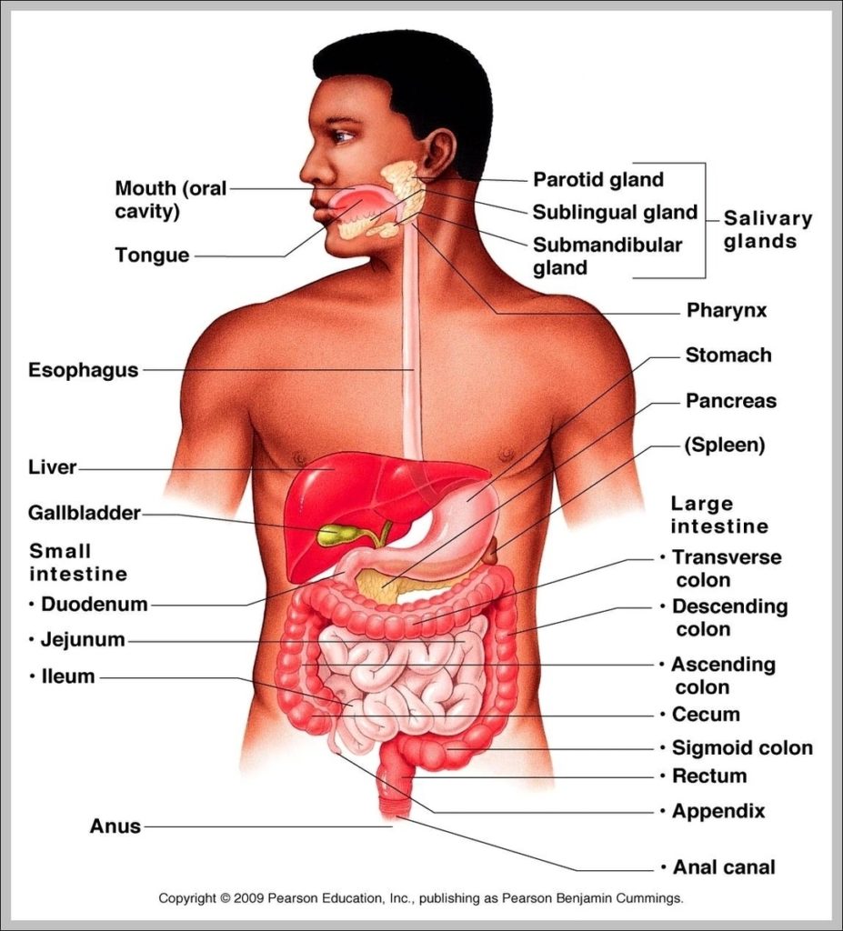
Your gallbladder is a small, pear-shaped organ in your upper right abdomen. Your gallbladder stores and releases bile to help your digestive system break down fats. The most common issue…
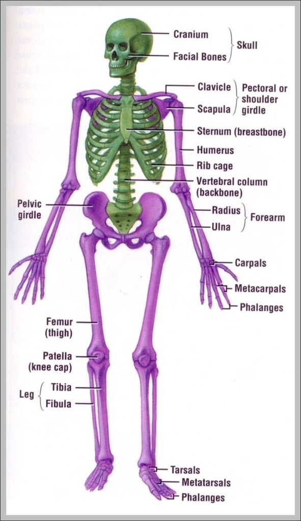
Human Organs The human body is the entire structure of a human being and comprises a head, neck, trunk (which includes the thorax and abdomen), two arms and hands and…
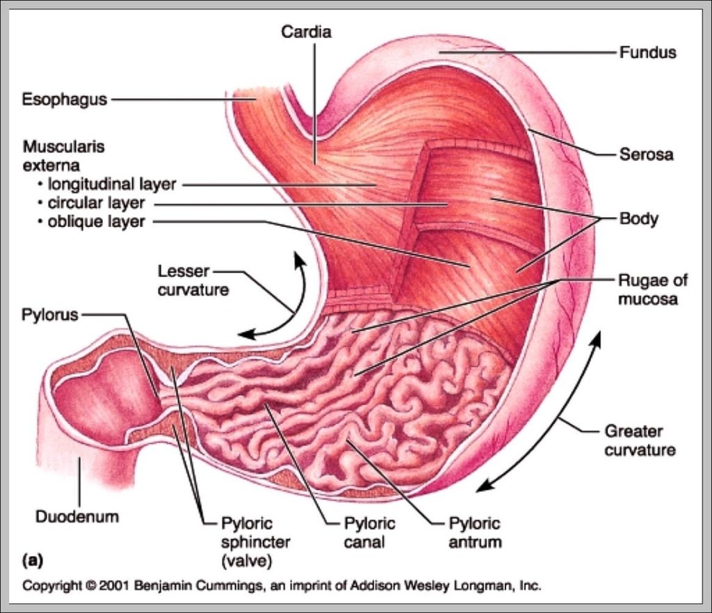
The size of the stomach varies from person to person. Your stomach expands when full and deflates when empty. Because of this, your stomach size can vary depending on how…
1,638 human head diagram stock photos and images available, or start a new search to explore more stock photos and images. An anatomical diagram consisting of a vertical cross-section of…
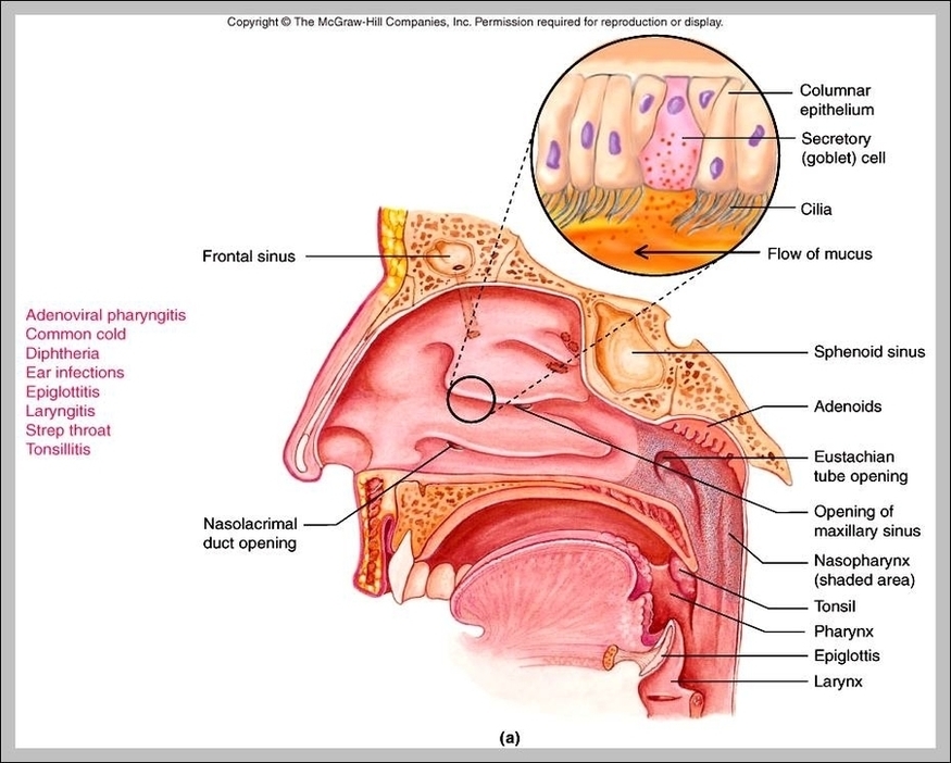
The main function of the epiglottis is to seal off the windpipe during eating, so that food is not accidentally inhaled. The epiglottis also helps with some aspects of sound…
Lymph node: Also sometimes referred to as lymph glands, lymph nodes are small rounded or bean-shaped masses of lymphatic tissue surrounded by a capsule of connective tissue. Lymph nodes are…
7,751 organs of the human body diagram stock illustrations and vector graphics available royalty-free, or start a new search to explore more great stock images and vector art. internal organs…
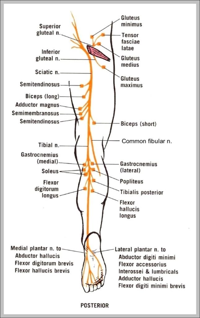
There are two main nerves in the leg: the femoral nerve serves the front and the sciatic nerve controls the back of the leg. The nerves of the leg can…
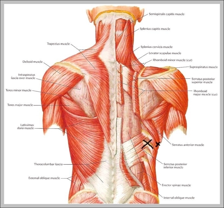
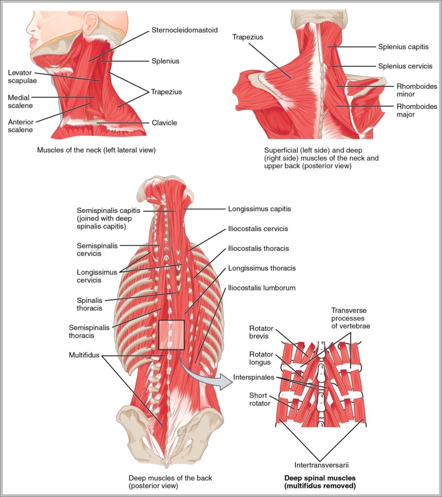
Muscles of neck. Neck muscles are bodies of tissue that produce motion in the neck when stimulated. The muscles of the neck run from the base of the skull to…
577 pics of the female buttocks anatomy stock photos and images available, or start a new search to explore more stock photos and images. Houman body parts flat line icons…
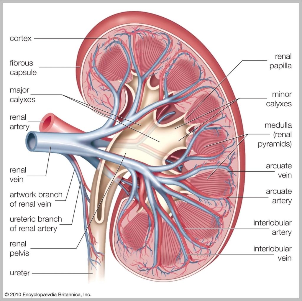
All rights reserved. Prev. Next. The kidneys are a pair of bean-shaped organs on either side of your spine, below your ribs and behind your belly. Each kidney is about…
77,785 nervous system stock photos and images available, or search for central nervous system or nervous system anatomy to find more great stock photos and pictures. Human nervous system medical…
Abstract image of a human body in the form of a starry sky or space, consisting of points, lines, and shapes in the form of planets, stars and the universe.…
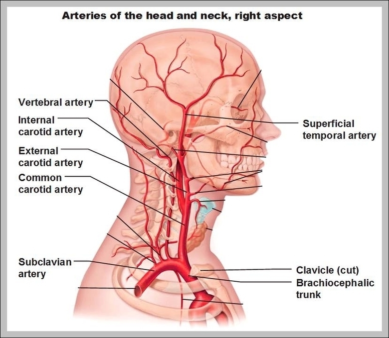
1,994 neck artery stock photos and images available, or search for head and neck artery to find more great stock photos and pictures. Photo Essay From Hospital. Photo Essay At…