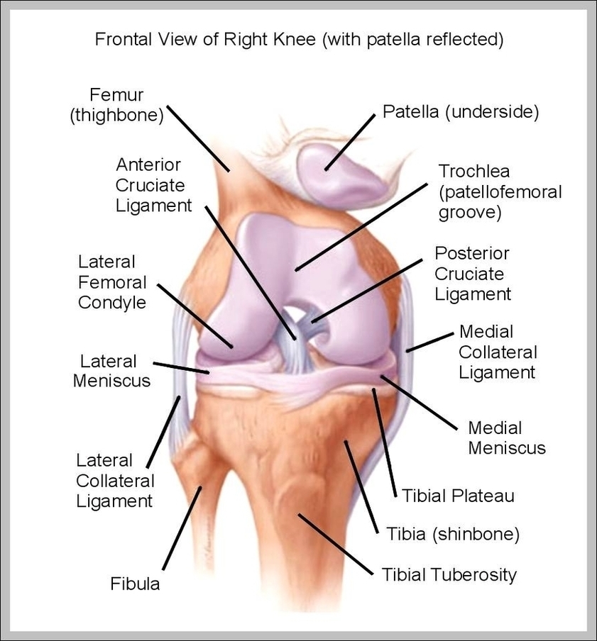
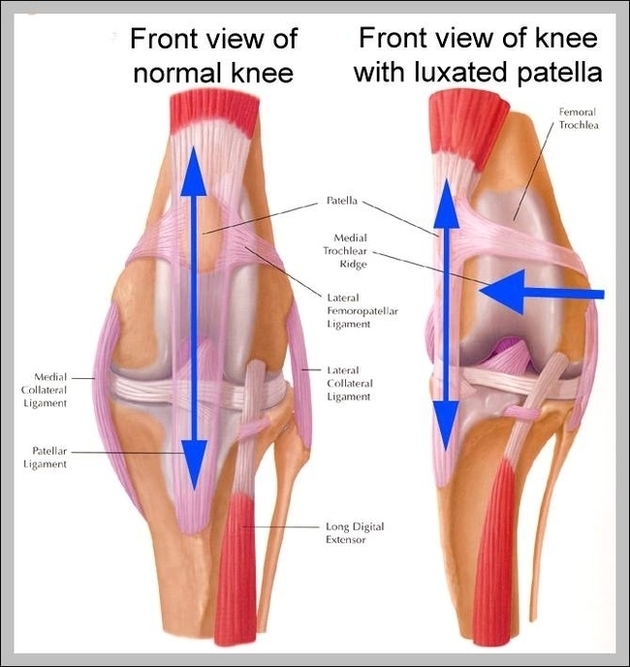
3,344 knee anatomy stock photos and images available, or search for knee anatomy illustration or human knee anatomy to find more great stock photos and pictures. To understand one of…
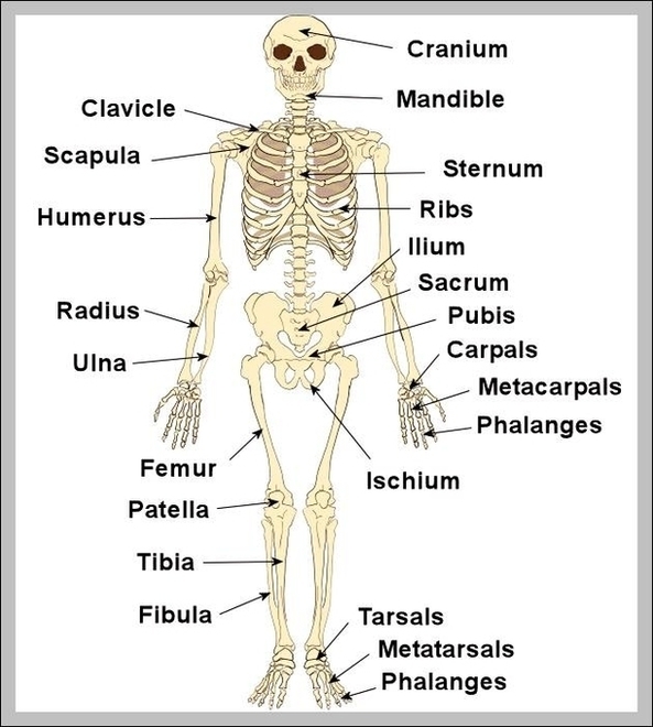
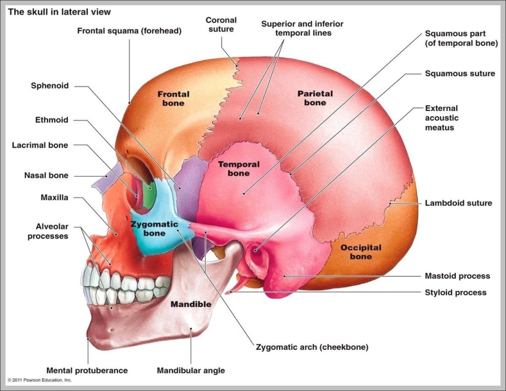
Save 25% when you use code SHARE25. 10,748 human skeleton labeled stock photos, vectors, and illustrations are available royalty-free. These bones are arranged into two major divisions: the axial skeleton…
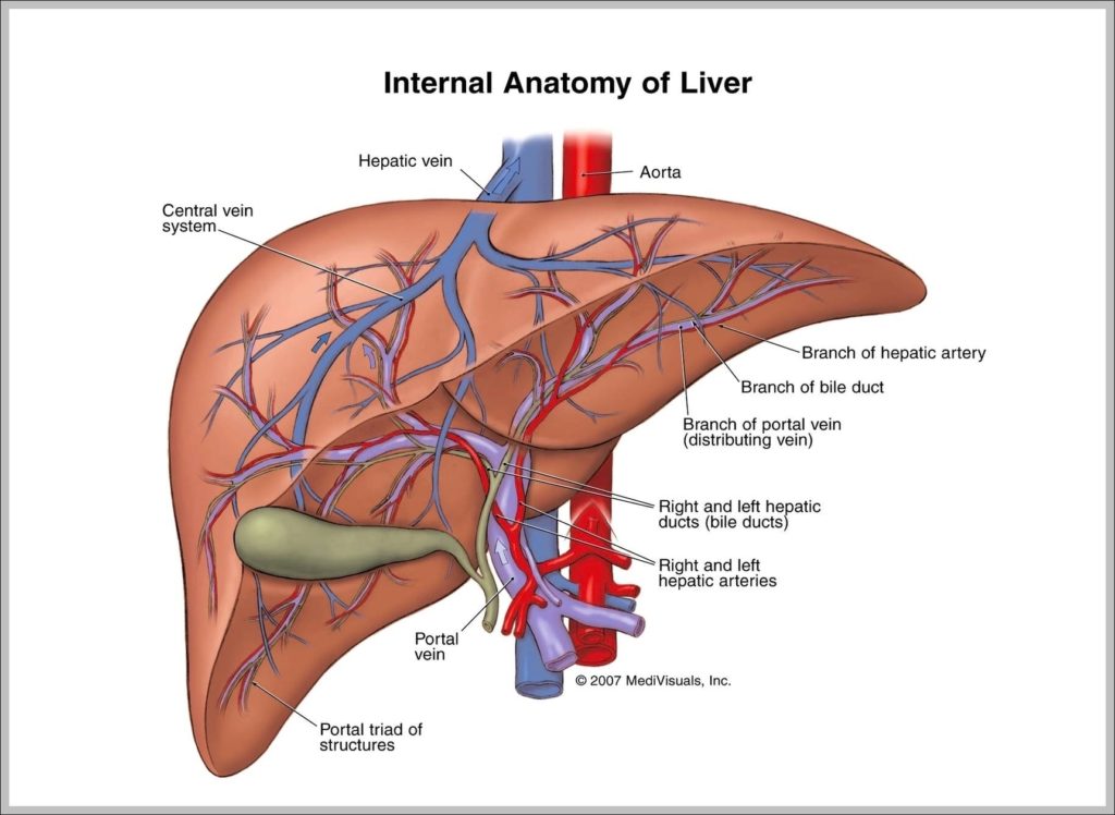
4,742 human liver anatomy stock photos and images available, or start a new search to explore more stock photos and images. The liver is a large, meaty organ that sits…
148,045 hand anatomy stock photos, vectors, and illustrations are available royalty-free. Picture of Hand. There are 28 phalanges (finger bones) and 10 metacarpal bones. Each finger has 3 phalanges and…
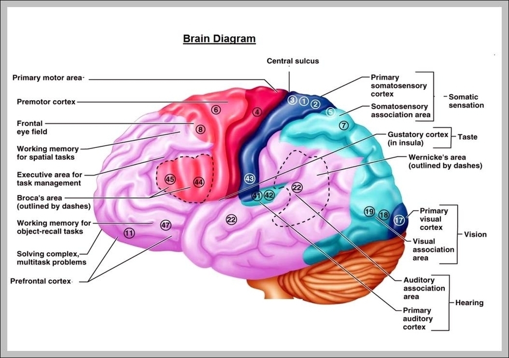
26,763 brain diagram stock photos, vectors, and illustrations are available royalty-free. This will help you understand the anatomy of the brain better. The average dimension of the adult human brain…
Light micrograph (bottom left) and computer illustration (top right) of the lining of the stomach, known as the mucosa. The stomach is a muscular sac involved in storage and digestion…
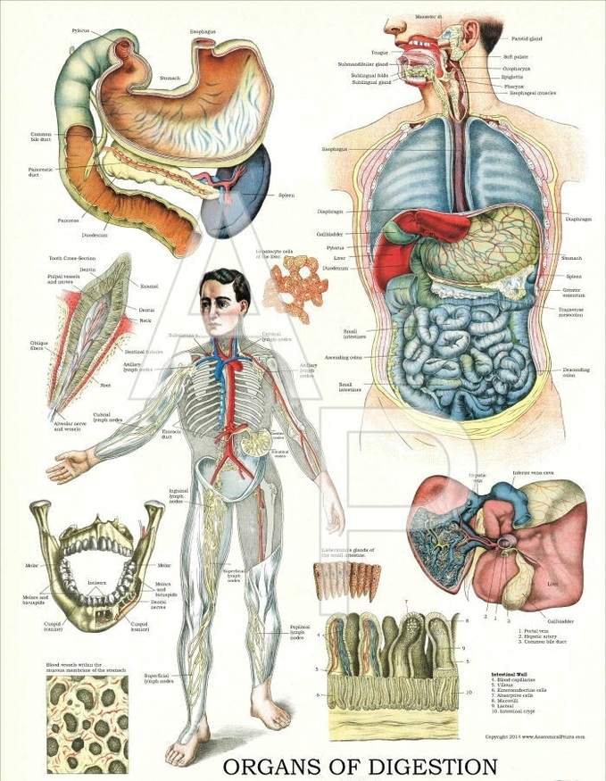
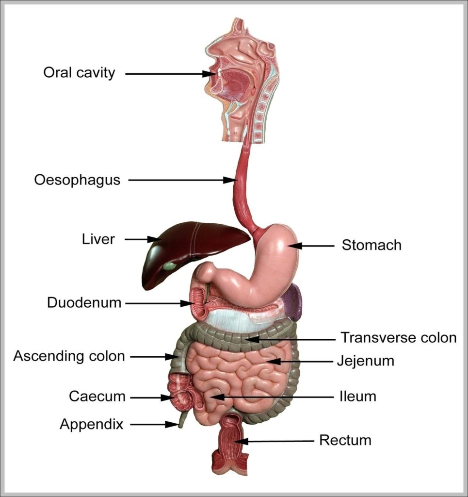
Liver –The liver is a large, reddish-brown, triangular-shaped organ of the digestive system, which is located to the right of the stomach. It functions by processing the absorbed food from…
717 image of pancreas in human body stock photos and images available, or start a new search to explore more stock photos and images. Anatomically, the pancreas is divided into…
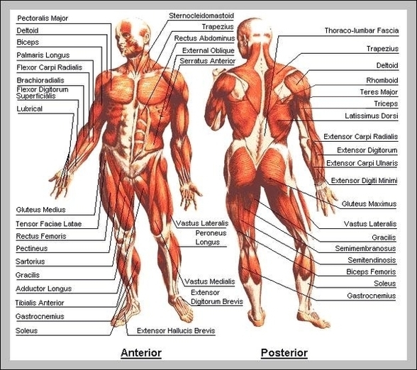
128,412 muscle anatomy stock photos, vectors, and illustrations are available royalty-free. We have included this interactive muscle map below to give you a visual guide to choose which parts of…
12,251 human body organs anatomy stock photos and images available, or start a new search to explore more stock photos and images. Human internal organs vector Vector isolated illustration of…
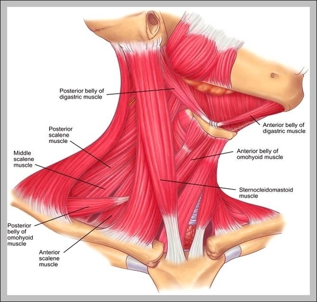
Common upper back pain symptoms include muscle stiffness, tightness, and tenderness that may affect the shoulders and neck. Upper back pain is often related to muscle or soft tissue problems,…
3,344 knee anatomy stock photos and images available, or search for knee anatomy illustration or human knee anatomy to find more great stock photos and pictures. Bad knee Images and…
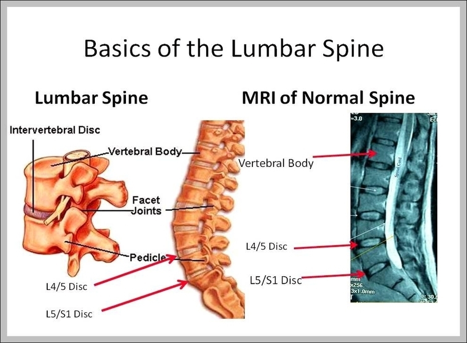
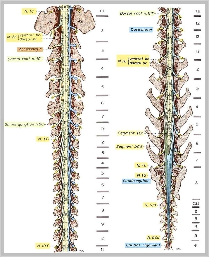
lumbar spine images 19,355 lumbar spine stock photos, vectors, and illustrations are available royalty-free. See lumbar spine stock video clips of 194 vertebrae illustrationintervertebral discscalciumvertebrevertebra structurelumbar vertebravertebral bodyhuman vertebraelumbarhip nerve…
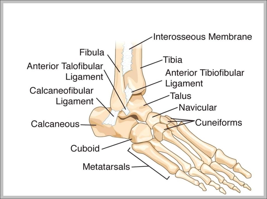
1,858 ankle bones stock photos and images available, or search for foot bones or knee to find more great stock photos and pictures. This makes the ankle one of the…
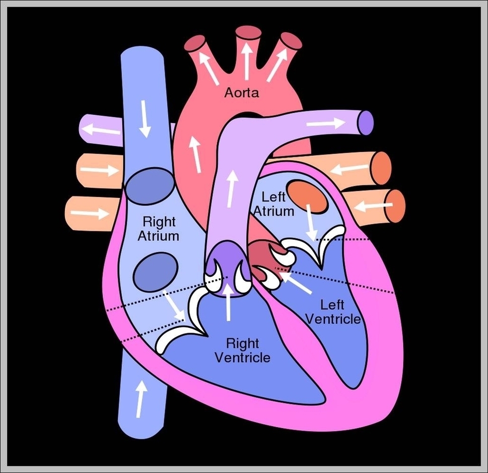
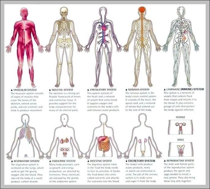
The 12 Animal Organ Systems. 1 The Respiratory System. SCIEPRO/Getty Images. All cells need oxygen, the crucial ingredient for extracting energy from organic compounds. Animals ... 2 The Circulatory System.…
The gastrointestinal tract is an organ system that enables us to ingest food via the mouth, digest it by breaking it down, absorb it, and then expel the remaining waste…