Cervical Spine Tinnitus Cervical spine tinnitus is a specific type of tinnitus where the perceived noises are believed to originate from disorders or dysfunctions in the cervical spine. The cervical…
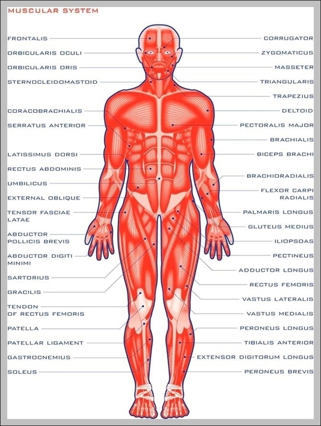
The strong muscles that move the ankle originate in the leg. The most well-known are the gastrocnemius and soleus muscles, which are the strong calf muscles that allow you to…
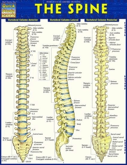
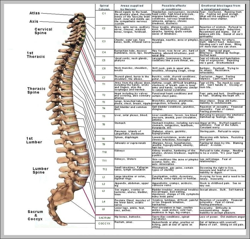
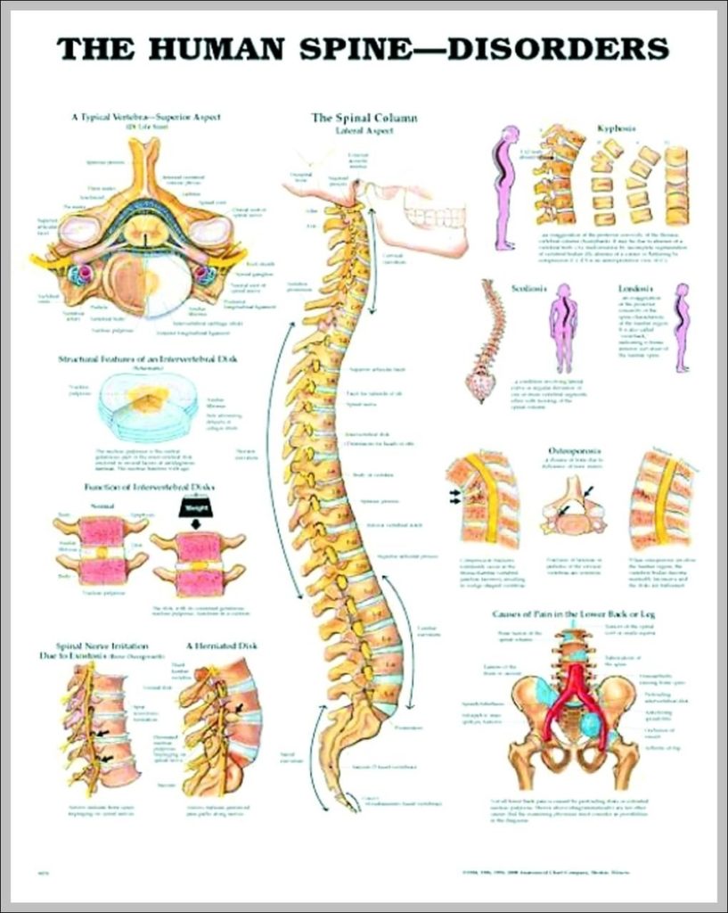
Your back consists of a complex array of bones, discs, nerves, joints, and muscles. The muscles of your back support your spine, attach your pelvis and shoulders to your trunk,…
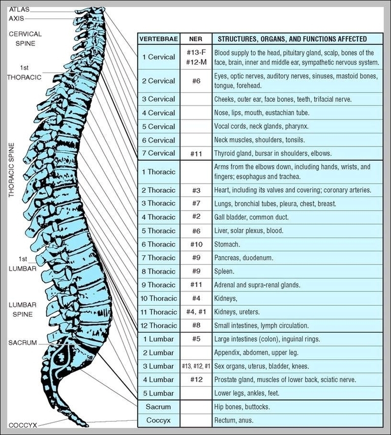
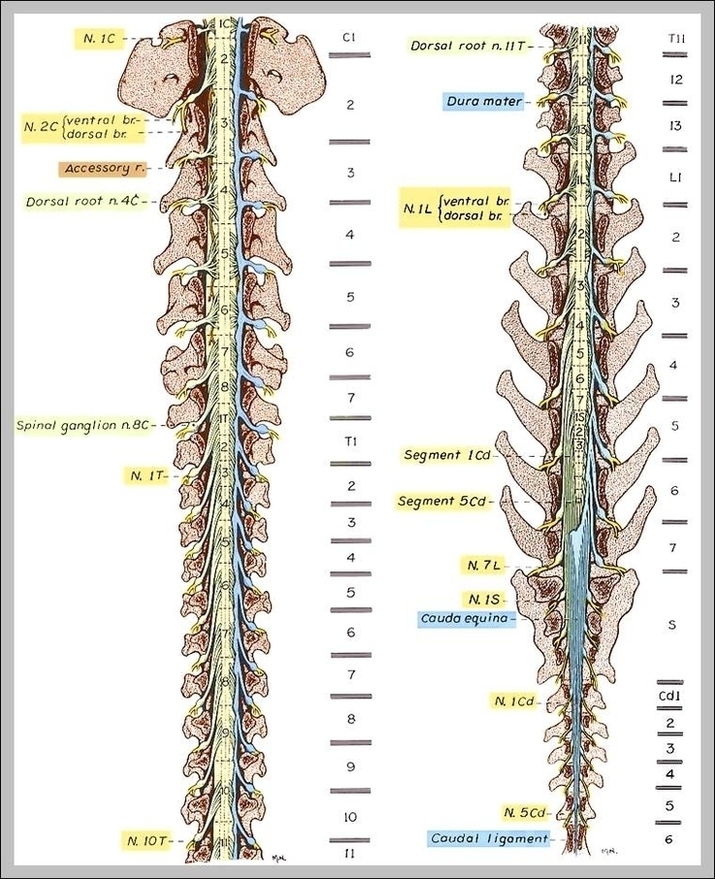
163,927 spine stock photos and images available, or search for back pain or spine xray to find more great stock photos and pictures. Human spine anatomy. Spinal segments and roots.…
21,076 human spine stock photos and images available or search for human spine anatomy or human spine xray to find more great stock photos and pictures. Search Term Spine By:…
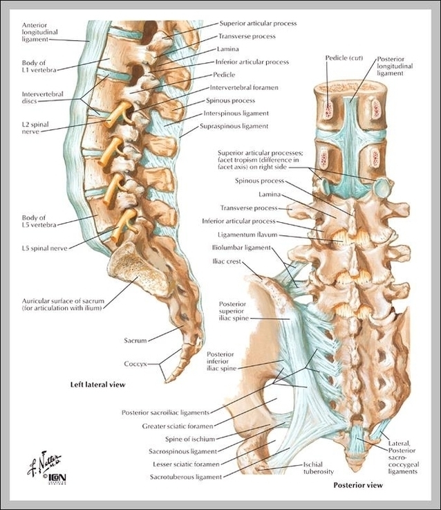
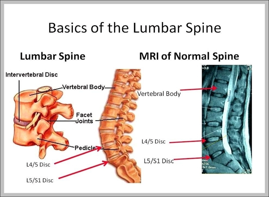
lumbar spine images 19,355 lumbar spine stock photos, vectors, and illustrations are available royalty-free. See lumbar spine stock video clips of 194 vertebrae illustrationintervertebral discscalciumvertebrevertebra structurelumbar vertebravertebral bodyhuman vertebraelumbarhip nerve…
Watch: Lumbar Spine Anatomy Video. "Lumbar" is derived from the Latin word "lumbus," meaning lion, and the lumbar spine earns its name. It is built for both power and flexibility…
lumbar spine images 19,355 lumbar spine stock photos, vectors, and illustrations are available royalty-free. See lumbar spine stock video clips of 194 vertebrae illustrationintervertebral discscalciumvertebrevertebra structurelumbar vertebravertebral bodyhuman vertebraelumbarhip nerve…
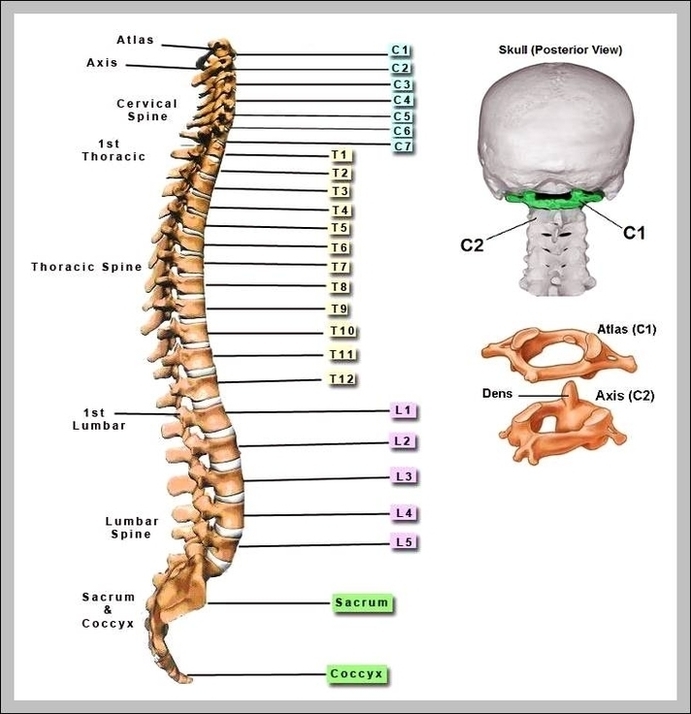
Toggle Anatomy System. Cervical vertebrae are the thinnest and most delicate vertebrae in the spine but offer great flexibility to the neck. The first cervical vertebra, C1, supports the skull…
All the images are in vector format, allowing an optimal web display with zoom and shifting of the anatomical images. A general view of the spine with the various levels…
21,109 human spine stock photos and images available, or search for human spine anatomy or human spine xray to find more great stock photos and pictures. Search Term Spine By:…
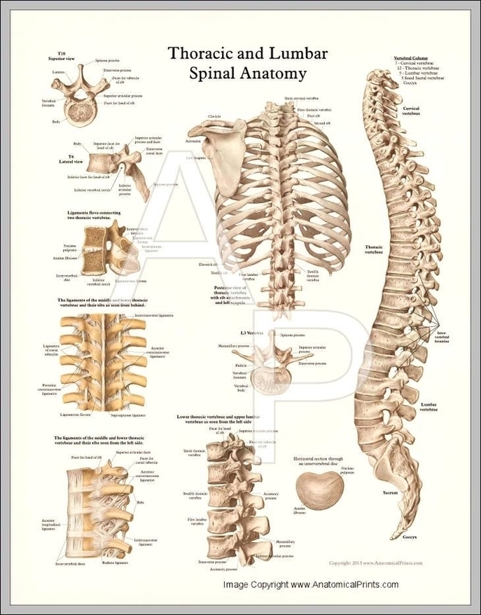
Anatomy of the Thoracic Spine. In technical terms, your spinal column at the mid and upper back levels is called the thoracic spine. The thoracic spine is comprised of 12…
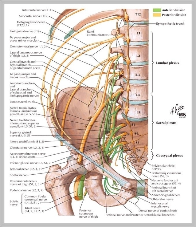
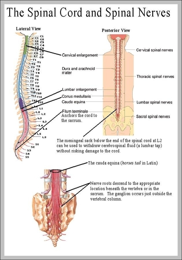
1,563 Spinal Nerve stock photos and images available, or search for spinal nerve block to find more great stock photos and pictures. Human nervous system medical vector illustration diagram with…
163,927 spine stock photos and images available, or search for back pain or spine xray to find more great stock photos and pictures. Human spine anatomy. Spinal segments and roots.…
Browse 21,109 human spine stock photos and images available, or search for human spine anatomy or human spine xray to find more great stock photos and pictures. Search Term Spine…
Spine Diagram A diagram of a human female spine showing a side view of the vertebra of the spinal cord within the the body. This is an editable EPS 10…