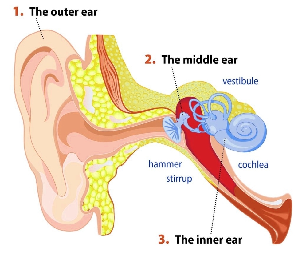
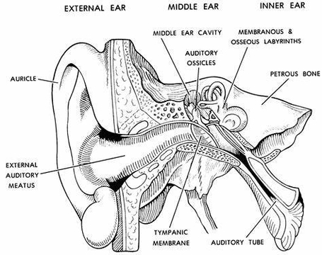
The inner ear, also known as the labyrinth, is the deepest part of the ear and plays a crucial role in hearing and maintaining balance. It consists of tiny bony…
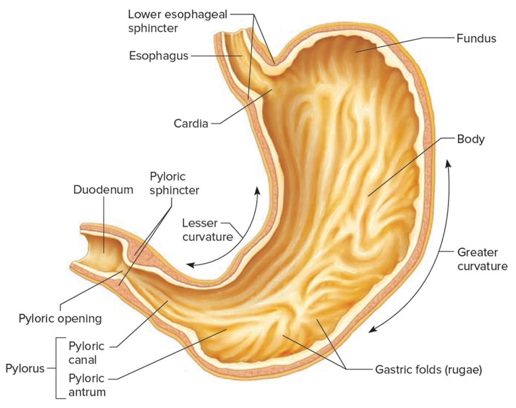
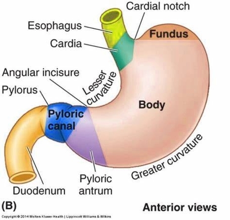
The stomach is a vital organ in the digestive system, responsible for breaking down food and sending it to the small intestine. It is located in the upper abdomen on…
The stomach is a key organ in the human digestive system, responsible for breaking down food and preparing it for further digestion and absorption in the intestines. It is located…
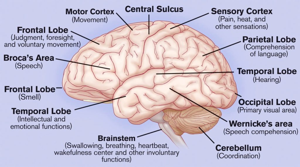
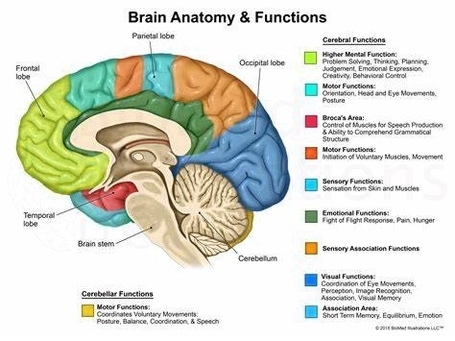
The human brain, an organ of immense complexity and the center of our consciousness, is divided into several distinct parts, each with its own specific functions. 1. Cerebrum The cerebrum,…
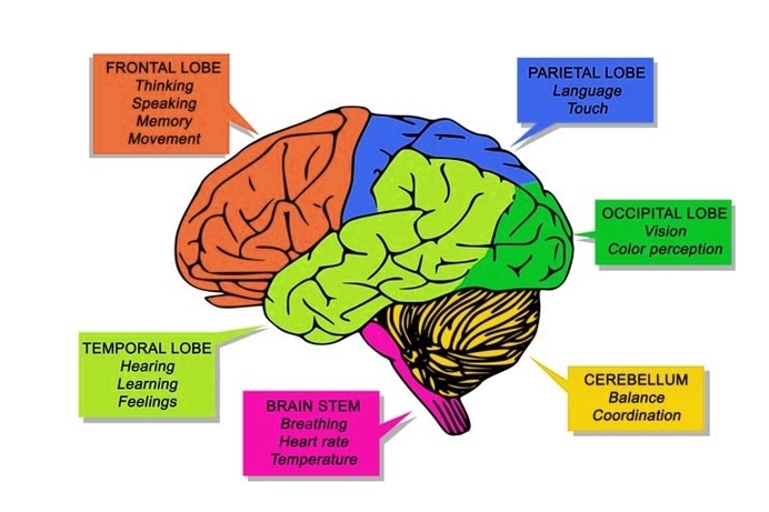
The human brain, the most complex organ in the body, is responsible for controlling thought, memory, emotion, touch, motor skills, vision, breathing, temperature, hunger, and every process that regulates our…
The human brain, the most complex organ in the body, is responsible for controlling thought, memory, emotion, touch, motor skills, vision, breathing, temperature, hunger, and every process that regulates our…
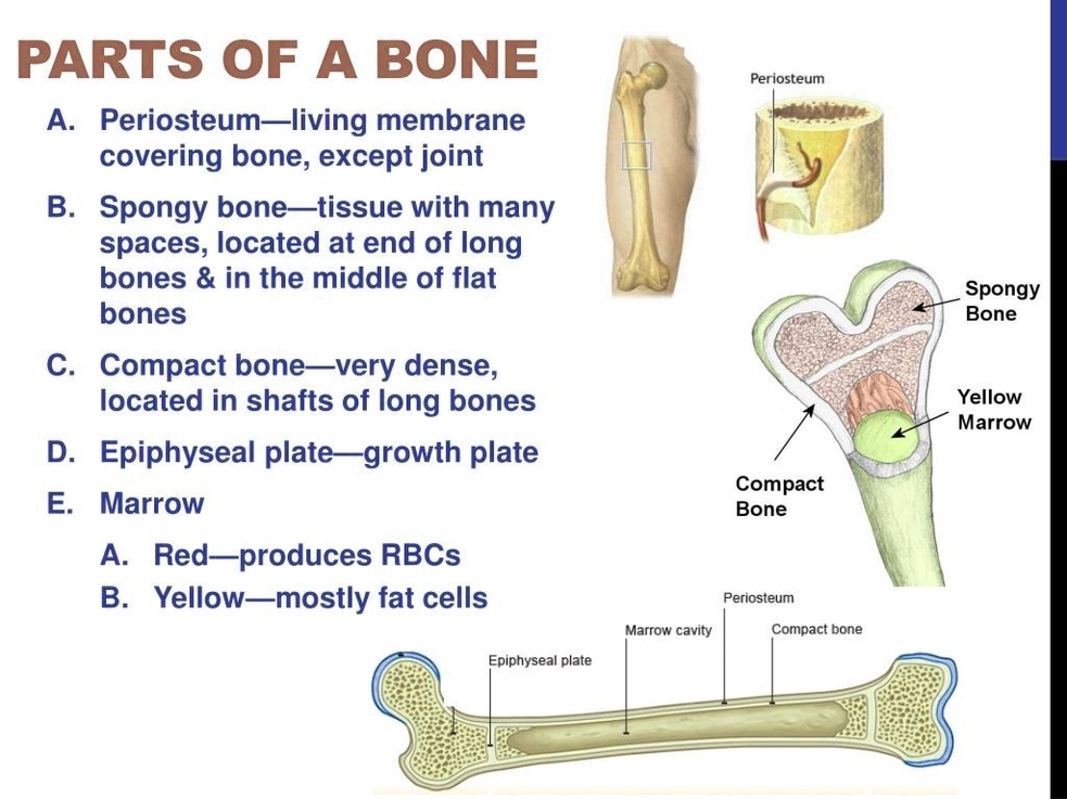
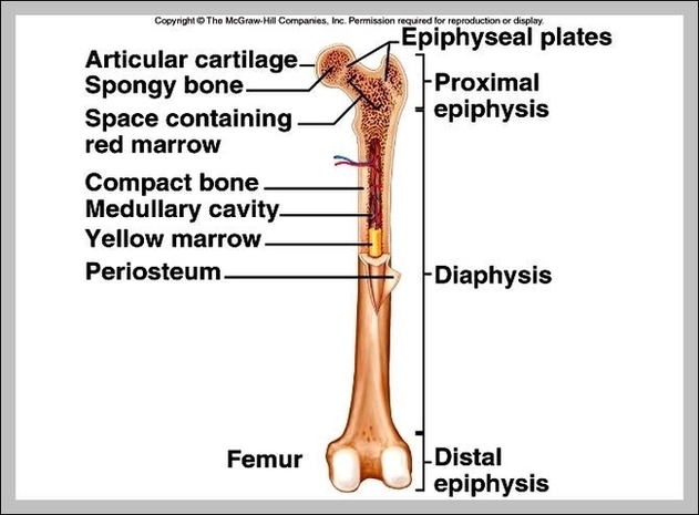
Bone Parts Bones are the structural framework of the body, providing support, protection, and facilitating movement. They are composed of several parts, each with a unique function: 1. Osteon: The…
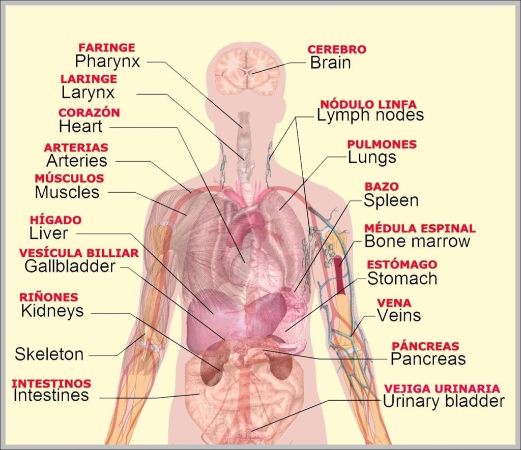
The names of body parts are used much the same as they are in Spanish as in English, but with one significant difference. In Spanish, names of parts of the…
Body parts for kids’ worksheets encourage the little ones to learn the primary names as they grow up. Keep the energy and eagerness for learning high with these fill in…
The parts of the small intestine are the: 1 Duodenum: The first and shortest section, which is roughly shaped like a "C." Food passes from... 2 Jejunum: Sugars, amino acids,…
2,101,867 male body parts stock photos and images available, or start a new search to explore more stock photos and images. Male External Genital Organs. See Images 0348407 For The…
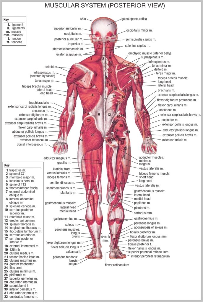
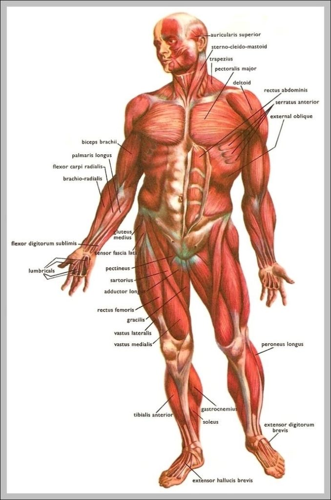
This muscular system picture shows all the major muscle groups on the human body from the frontal view. To see a muscular system diagram from the posterior (back) view click…
28,679 anatomy of the human body stock photos and images available, or start a new search to explore more stock photos and images. Human body systems. Illustrations of the muscular,…
respiratory system: bronchiole and bronchi, diaphragm, trachea, alveoli and cross-section of the lungs. Vector illustration for your design and medical use. human anatomy. silhouette of a man on white background.…
309 external human body parts stock photos, vectors, and illustrations are available royalty-free. 4,039,116 human body parts stock photos and images available, or search for human body parts icons or…
4,902 hip anatomy stock photos and images available, or search for human hip anatomy or hip anatomy illustration to find more great stock photos and pictures. Picture of Hip. The…
5,816 body parts diagram illustrations, drawings, and clip-art are available royalty-free. 7,751 organs of the human body diagram stock illustrations and vector graphics available royalty-free, or start a new search…