Lower Leg Muscles Study The lower leg, anatomically defined as the region of the lower limb below the knee, is a complex structure that plays a crucial role in movements…
Leg Muscles: An Overview The leg muscles, comprising several strong muscles in the upper and lower leg, play a crucial role in facilitating various movements, supporting body weight, and maintaining…
Anatomy of Leg Muscles The leg, anatomically defined as the region of the lower limb from the knee to the ankle, is composed of various muscles that enable movements like…
The lower leg, anatomically defined as the region of the lower limb below the knee, is a complex structure that plays a crucial role in weight-bearing activities such as walking,…
The human leg, anatomically defined as the region of the lower limb from the knee to the ankle, is a complex structure composed of numerous muscles that work in harmony…
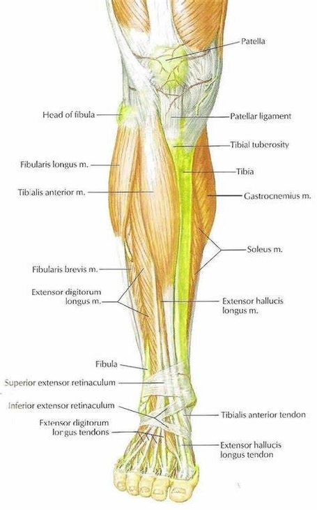
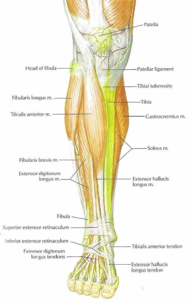
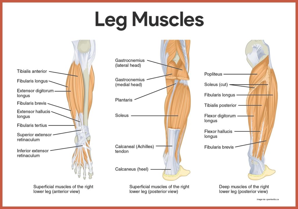
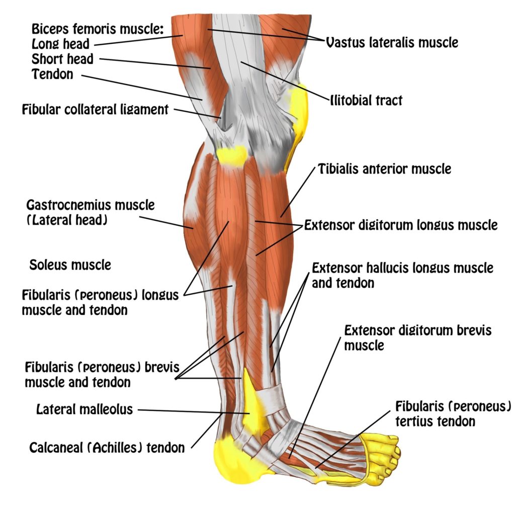
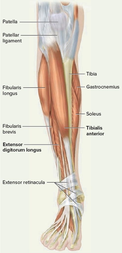
the anterior compartment muscles of the leg. These muscles play a crucial role in movement, stability, and overall function of the lower limb. Without further ado, let's explore their anatomy,…
Understanding Leg Muscles and Their Importance in Gym Workouts The leg muscles are a complex group of muscles that not only support the body's weight but also play a crucial…
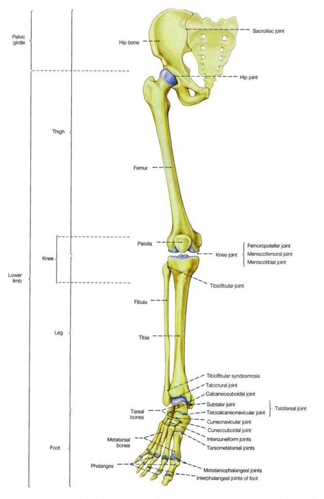
The human leg, a marvel of biological engineering, is a complex structure composed of numerous bones. These bones are designed to withstand daily strain, absorb force, and provide flexibility for…
The human leg, a marvel of biological engineering, is composed of numerous bones that provide structure, mobility, and support to the body. These bones, from the hip down to the…
The lower leg, anatomically defined as the region of the lower limb below the knee, is a complex structure that plays a crucial role in movements such as walking, running,…
The lower leg, also known as the "leg" in anatomical terms, is a significant part of the skeletal system. It forms the lower extremity along with the upper leg and…
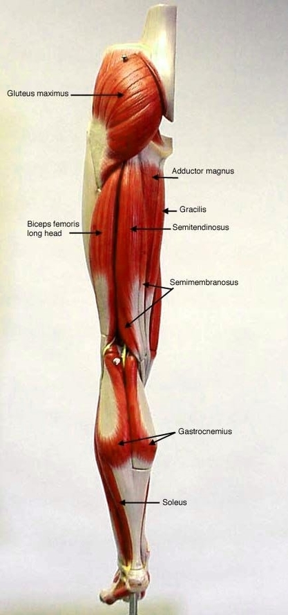
The human leg, a marvel of biological engineering, is powered by a complex network of muscles. These muscles, working in harmony, enable us to perform a wide range of movements,…
Leg Muscles Anatomy Study The leg muscles, anatomically defined as the region of the lower limb below the knee, are organized into three compartments: anterior, posterior, and lateral. Anterior (Dorsiflexor)…
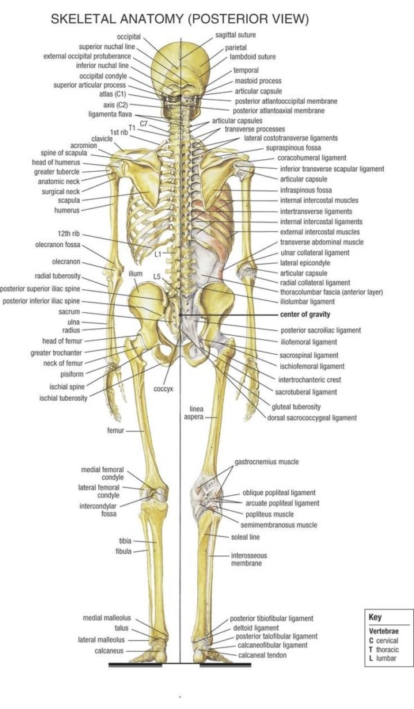
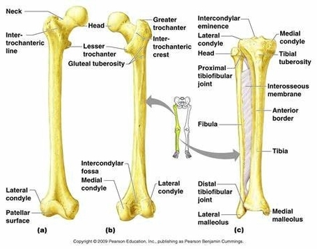
The human leg is a complex structure composed of various bones that play a crucial role in movement, weight-bearing, and maintaining balance. Here's a detailed look at the bones that…
Human Leg Bones: Labeled, Defined, and Described The human leg, a marvel of biological engineering, is a complex structure composed of numerous bones. These bones, from the hip down to…
The human leg, a marvel of biological engineering, is a complex structure composed of numerous bones that work in harmony to provide support and mobility. Femur (Thighbone) The femur, or…
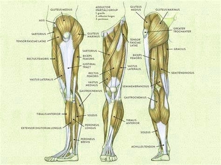
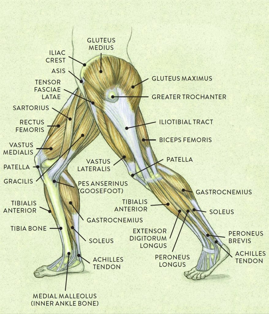
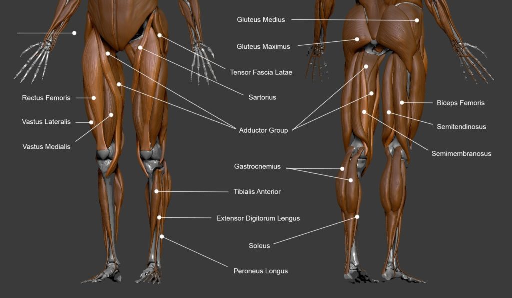
The human leg, a complex structure that enables movement and supports the body's weight, is composed of numerous muscles. These muscles can be broadly categorized into those of the upper…
Quadriceps Femoris: The Powerhouse of the Leg The quadriceps femoris, commonly known as the quads, is a group of muscles located at the front of the thigh. They are some…
Leg Muscles and Ligaments The leg, anatomically defined as the region of the lower limb below the knee, is a complex structure that includes various muscles and ligaments. These components…
Tendons are thick bands of tissue that connect muscles to bone. When a muscle contracts, the tendon pulls on the bone causing the joint to move. There are a number…