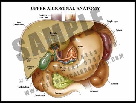
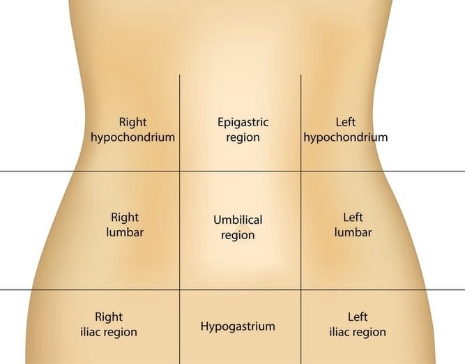
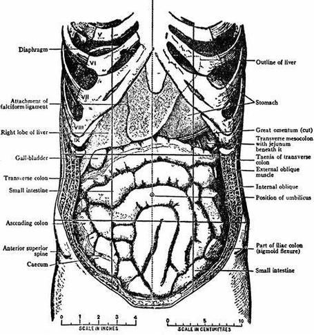
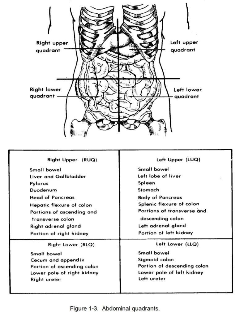
Abdominal Anatomy Picture Image

The main function of the epiglottis is to seal off the windpipe during eating, so that food is not accidentally inhaled. The epiglottis also helps with some aspects of sound production in certain languages.
Epiglottis – dorsal view. The epiglottis is one of the three large unpaired laryngeal cartilages, the other two being the book shaped thyroid and the signet ring shaped cricoid cartilages. The thyroid notch is also termed the laryngeal prominence/Adam’s apple.

Abdominal Anatomy Picture Image Diagram - Abdominal Anatomy Picture Image Chart - Human anatomy diagrams and charts explained. This anatomy system diagram depicts Abdominal Anatomy Picture Image with parts and labels. Best diagram to help learn about health, human body and medicine.

Abdominal Anatomy Picture Image

Abdominal Arteries Image

The aorta is the largest blood vessel in the body. It is an artery, meaning that it carries blood away from the heart. The abdominal aorta enters the abdomen through the diaphragm at the level of the twelfth thoracic vertebre and continues to just below the umbilical area, where it splits into the right and left common iliac arteries.
The abdominal arteries include: Celiac trunk. Branching off from the abdominal aorta, the celiac trunk divides into smaller arteries that supply organs such as the stomach, liver, and spleen. Superior mesenteric.

Abdominal Arteries Image Diagram - Abdominal Arteries Image Chart - Human anatomy diagrams and charts explained. This anatomy system diagram depicts Abdominal Arteries Image with parts and labels. Best diagram to help learn about health, human body and medicine.

Abdominal Arteries Image

Internal Abdominal Oblique Image

The abdominal internal oblique muscle, also internal oblique muscle or interior oblique, is an abdominal muscle in the abdominal wall that lies below the external oblique muscle and just above the transverse abdominal muscle . The abdominal internal oblique muscle. Muscles of the trunk.
(T12), Iliohypogastric n. (L1) and Ilioinguinal n. (L1) The abdominal internal oblique muscle, also internal oblique muscle or interior oblique, is an abdominal muscle in the abdominal wall that lies below the external oblique muscle and just above the transverse abdominal muscle .

Internal Abdominal Oblique Image Diagram - Internal Abdominal Oblique Image Chart - Human anatomy diagrams and charts explained. This anatomy system diagram depicts Internal Abdominal Oblique Image with parts and labels. Best diagram to help learn about health, human body and medicine.

Internal Abdominal Oblique Image

Abdominal Regions

Each of the 9 abdomen regions hold their own important organs: Right upper quadrant – This will be assessed by doctors for tenderness and also localised pain from organs such as the gall bladder, liver, colon (hepatic flexure), duodenum, and the upper part of the pancreas.

Region 9 is known as the left iliac fossa region, home to the descending colon. There are nine regions of the abdomen that house various organs. These regions are broken into three areas: the upper, middle, and lower abdomen. In the upper abdomen, the right hypochondriac, epigastric, and left hypochondriac regions reside.

Please try again later. Here are some of the major organs that you’ll find in each of the four abdominal quadrants: Right Upper Quadrant: Liver, stomach, gallbladder, duodenum, right kidney, pancreas, and the right adrenal gland. Left Upper Quadrant: Liver, stomach, pancreas, left kidney, spleen, and the left adrenal gland.

Abdominal Regions Diagram - Abdominal Regions Chart - Human anatomy diagrams and charts explained. This anatomy system diagram depicts Abdominal Regions with parts and labels. Best diagram to help learn about health, human body and medicine.

abdominal regions