Inner Ear Anatomy
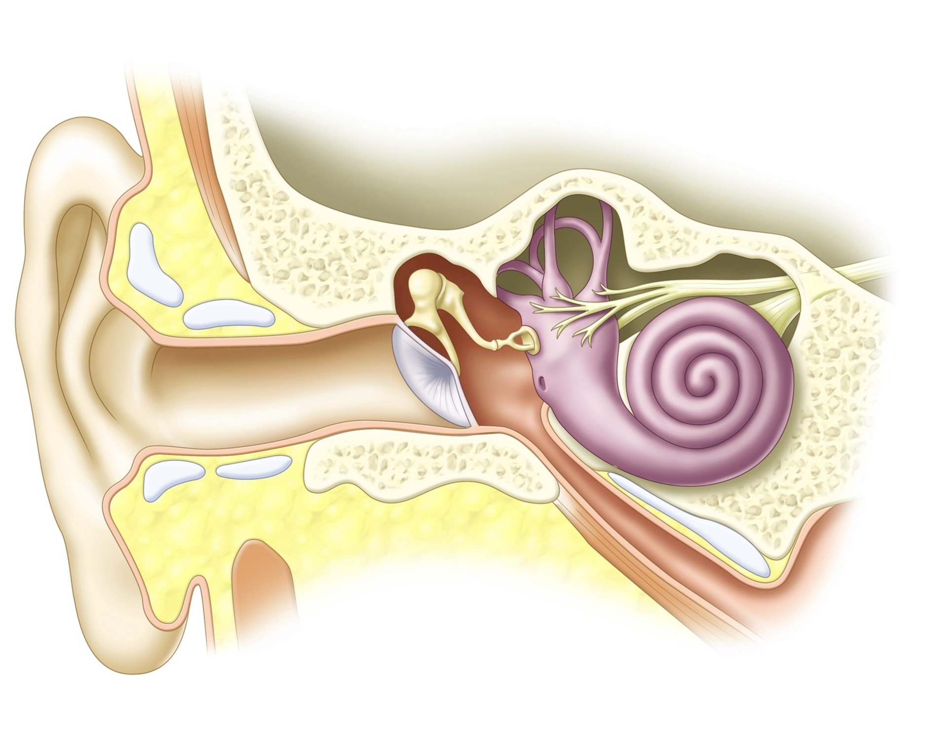
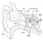
The inner ear, also known as the labyrinth, is a complex structure responsible for hearing and balance. It is the innermost part of the ear and consists of tiny bony structures filled with fluid.
Components of the Inner Ear
The inner ear comprises the bony labyrinth and the membranous labyrinth.
1. Bony Labyrinth: This includes the cochlea, semicircular canals, and vestibule.
– Cochlea: Shaped like a snail, the cochlea is divided into two chambers by a membrane. These chambers are filled with fluid, which vibrates when sound enters, causing the tiny hairs lining the membrane to vibrate and send electrical impulses (sound signals) to the brain.
– Semicircular Canals: Also known as the labyrinthine, these canals rest on top of the cochlea, connected by the vestibule. They are filled with fluid and contain small calcium crystals and tiny hairs that sense the movement of the fluid.
– Vestibule: The vestibule is the central part of the bony labyrinth. It communicates anteriorly with the cochlea and posteriorly with the semicircular canals.
2. Membranous Labyrinth: This lies inside the bony labyrinth and also consists of three parts.
– Cochlear Duct: This triangle-shaped duct


