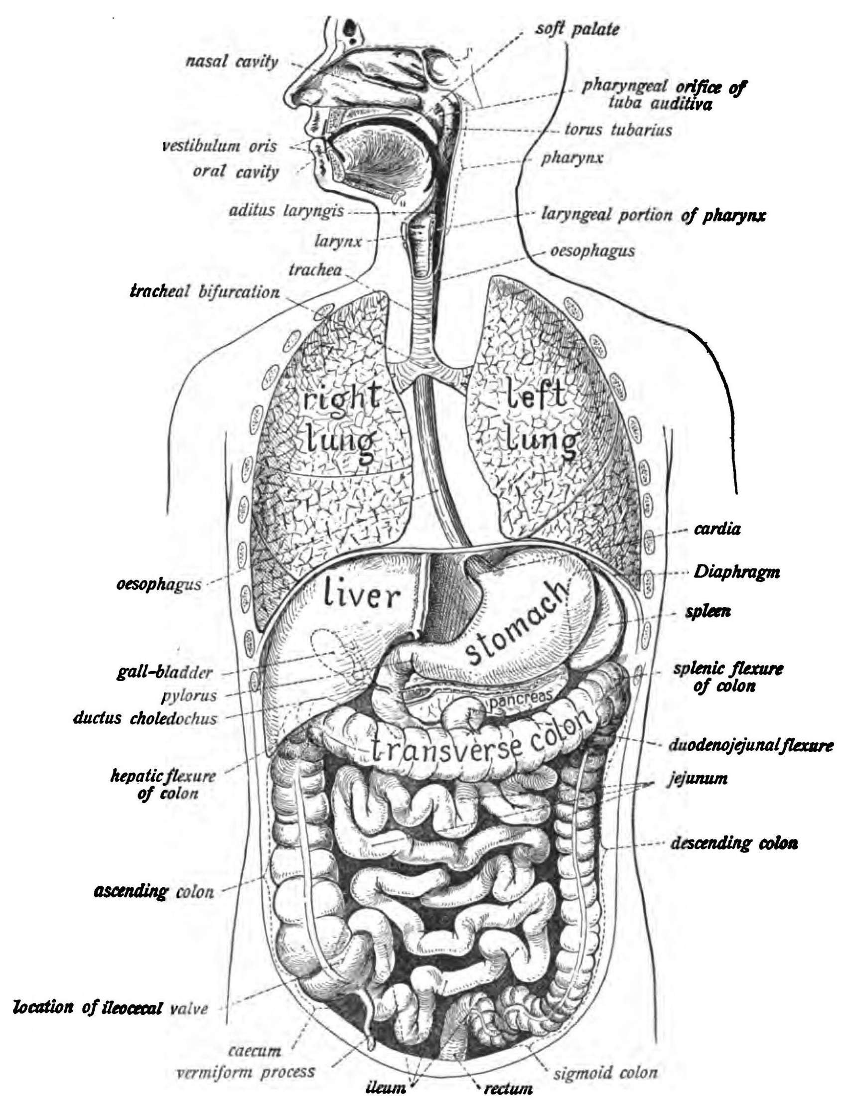
abdominal organs. These vital structures play a crucial role in digestion, metabolism, and overall well-being. While I won’t provide visual images, I’ll paint a vivid picture with words.
1. Liver:
– The liver, our body’s largest internal organ, resides in the upper right quadrant of the abdomen. It acts as a multifunctional powerhouse.
– Functions:
– Detoxification: The liver filters toxins and waste products from the blood.
– Bile Production: It produces bile, essential for fat digestion.
– Storage: The liver stores nutrients like vitamins and glycogen.
– Beneath the liver lies the gallbladder, a small sac that stores excess bile until needed for digestion.
2. Stomach:
– The stomach sits just below the liver. It’s a muscular pouch where food enters after swallowing.
– Functions:
– Storage: The stomach temporarily holds food.
– Mixing and Churning: Muscles contract to mix food with digestive juices.
– Acid Secretion: Hydrochloric acid aids in breaking down proteins.
– Pepsin: Enzymes like pepsin further digest proteins.
3. Pancreas:
– The pancreas lies behind the stomach, resembling a tadpole.
– Functions:
– Enzyme Production: It produces enzymes (amylase, lipase, protease) for digesting carbohydrates, fats, and proteins.
– Hormone Regulation: The pancreas secretes insulin and glucagon, regulating blood sugar levels.
4. Small Intestine:
– The small intestine, a remarkable 21-foot-long tube, dominates the abdominal cavity.
– Functions:
– Digestion and Absorption: Here, fats, starches, and proteins break down into absorbable nutrients.
– Villi and Microvilli: These tiny finger-like projections increase surface area for nutrient absorption.
5. Large Intestine (Colon):
– The large intestine follows the small intestine, though it’s shorter but wider.
– Functions:
– Water Absorption: It reabsorbs water from undigested food.
– Fermentation: Beneficial gut bacteria ferment undigested carbohydrates.
– Formation of Feces: The colon compacts waste into feces.
6. Kidneys:
– The kidneys, located behind the intestines, are essential for filtering blood.
– Functions:
– Nephrons: Each kidney contains about a million nephrons that filter blood and regulate electrolytes.
– Urine Formation: Excess waste and water form urine, which drains into the bladder.
7. Adrenal Glands:
– Atop each kidney sit the adrenal glands.
– Functions:
– Cortisol and Adrenaline: These glands produce hormones like cortisol (stress response) and adrenaline (fight-or-flight).
8. Ureters:
– The ureters are slender tubes connecting the kidneys to the bladder.
– Functions:
– They transport urine from the kidneys to the bladder.
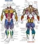
9. Ribs and Rib Cage:
– The rib cage encloses and protects these organs.
– Functions:
– The ribs shield vital structures from external trauma.
– The sternum (breastbone) anchors the upper ribs.
In summary, the abdominal organs orchestrate a symphony of functions, ensuring our survival and well-being. From digestion to waste elimination, they work tirelessly behind the scenes, allowing us to savor life’s flavors and thrive. ????
For further exploration, consider medical illustrations or 3D models that vividly depict these intricate structures ..
o