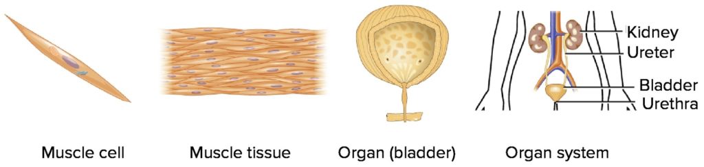
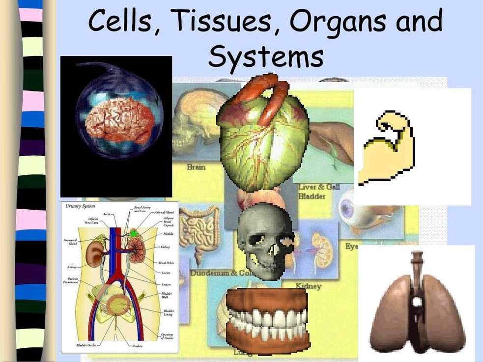
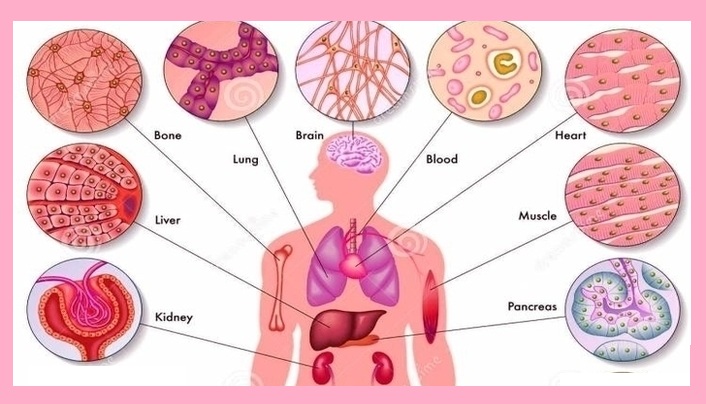
Cells, Tissues, Organs, and Systems Cells are the smallest unit of life. They come in various shapes and serve specialized purposes in the body. For instance, muscle cells contract, nerve…
Cells Cells are the basic building blocks of all living things?. They provide structure for the body, take in nutrients from food, convert those nutrients into energy, and carry out…
The digestive system, a complex network of organs, plays a crucial role in the body's overall health and functioning. It is responsible for breaking down food into nutrients, which the…
5,816 body parts diagram illustrations, drawings, and clip-art are available royalty-free. 7,751 organs of the human body diagram stock illustrations and vector graphics available royalty-free, or start a new search…
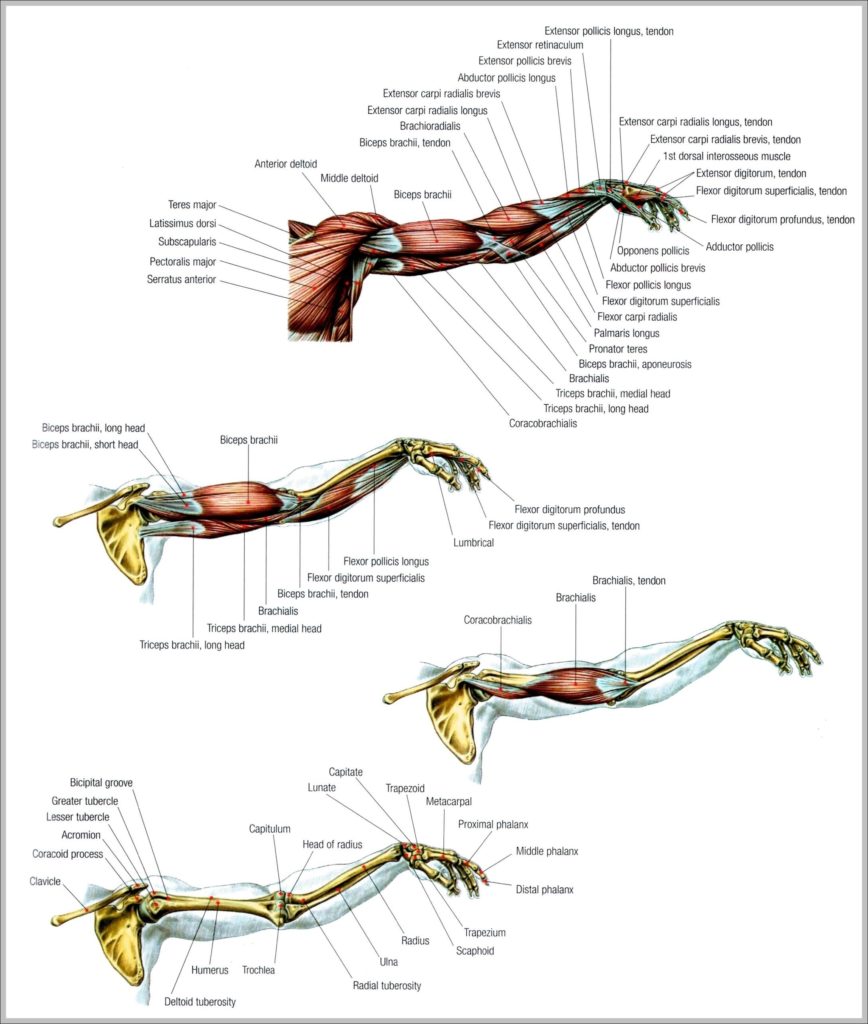
Muscle Cell Types 1 Skeletal Muscle. Skeletal muscle is the most common and widely distributed muscle tissue in the body, making up around 40% of the body’s total mass. 2…
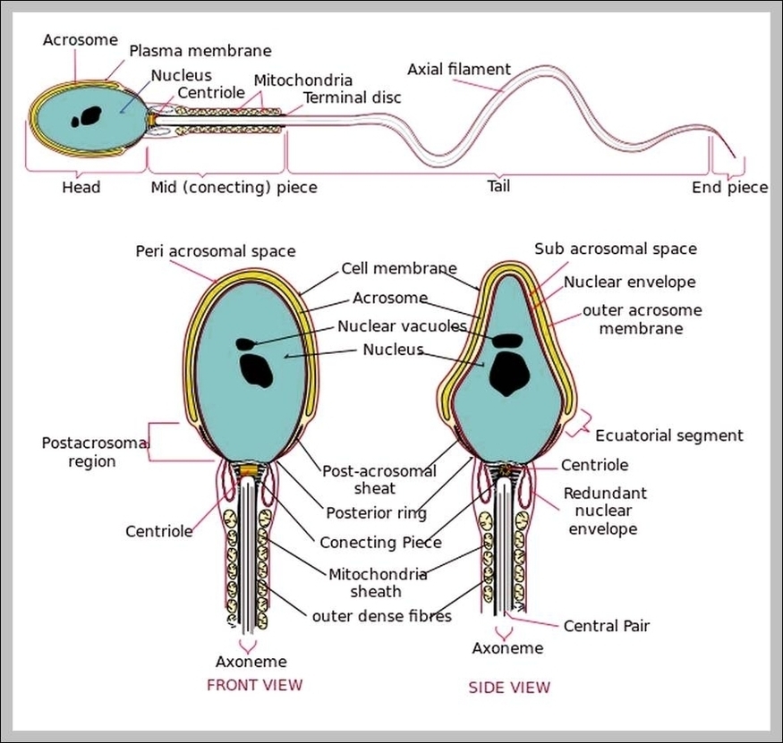
Sperm cells are gametes (sex cells) that are produced in the testicular organ (gonad) of male human beings and animals. Like the female gamete (oocyte), sperm cells carry a total…
1,416 muscle cell stock photos and images available, or search for smooth muscle cell or skeletal muscle cell to find more great stock photos and pictures. Sem Of Muscle Fiber.…
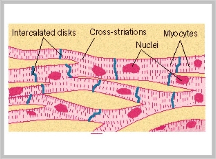
2,803 cardiac muscle cell stock photos and images available, or start a new search to explore more stock photos and images. As the chief cell type of the heart, cardiac…
Stomach histology. The stomach is a key part of the gastrointestinal (GI) tract, sitting between the esophagus and duodenum. Its functions are to mix food with stomach acid and break…
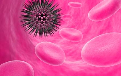
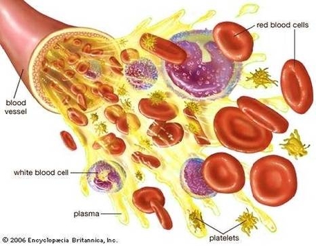
Cancer develops when cells in the body multiply out of control. Blood contains three kinds of cells: red cells, white cells, and platelets. Any of these kinds of cells can…
Diagram of the human cell illustrating the different parts of the cell. The cell membrane is the outer coating of the cell and contains the cytoplasm, substances within it and…
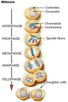
Mitosis. process cell division. Mitosis is the process by which our bodies replace cells. Daughter cells have identical chromosomes to parent cell, genetic Diagram of the Cell Cycle. Types of…
Red blood cells (RBC) which transport oxygen to cells of the body White blood cells (WBC), which are a part of the immune system Hemoglobin (Hb), a protein which carries…
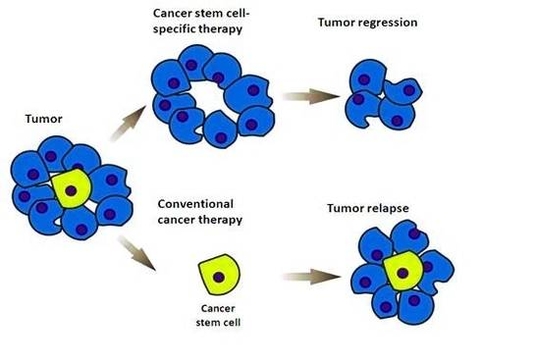
Cancer stem cells ( CSCs) are cancer cells (found within tumors or hematological cancers) that possess characteristics associated with normal stem cells, specifically the ability to give rise to all…
1,950 red blood cell diagram stock photos, vectors, and illustrations are available royalty-free. Blood cells are the cells which are produced during hematopoiesis and found mainly in the blood. Blood…
Human Cell Diagram, Parts, Pictures, Structure and Functions. The cell is the basic functional in a human meaning that it is a self-contained and fully operational living entity. Humans are…
A detailed explanation of the heart along with a well-labelled diagram is given for reference. The upper two chambers of the heart are called auricles. The lower two chambers of…