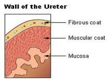
The elementary structure of the ureter is elastic muscles entangled in fiber layers, that allow to control the sphincter. The muscular layers cover the whole path between the kidney to the bladder. The kidneys produce urine by filtering excess water from our blood. The blood transports the debris to the kidneys.
From a histological perspective, there are two muscular layers in the wall of the ureter: a longitudinal and a circular layer. In the lower segment of the ureters, another longitudinal layer can be found proximal to the bladder.
The ureters measure between 20 and 26 cm. The muscles in the ureters walls contract and relax, in order to force the urine to go away from the kidneys. Small quantities of urine flow from the ureters to the bladder every single 10-15 seconds. Through a sequences of ureters walls contractions and relaxations, the tubular structure advances.

ureters wall diagram
Posted inDiagrams
