Muscles Of Facial Expression Image

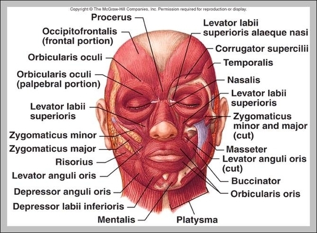
The specific location and attachments of the facial muscles enable them to produce movements  of the face, such as smiling, grinning and frowning. Thus, these muscles are commonly called muscles of facial expression, or mimetic muscles. All of the facial muscles are innervated by the facial nerve (CN VII)  and vascularized by the facial artery .
This field is for validation purposes and should be left unchanged. The muscles of facial expression (also known as the mimetic muscles) can generally be divided into three main functional categories: orbital, nasal and oral. These muscles are all innervated by the facial nerve (CN VII).¹

Muscles Of Facial Expression Image Diagram - Muscles Of Facial Expression Image Chart - Human anatomy diagrams and charts explained. This anatomy system diagram depicts Muscles Of Facial Expression Image with parts and labels. Best diagram to help learn about health, human body and medicine.

Muscles Of Facial Expression Image

Facial Bones Diagram

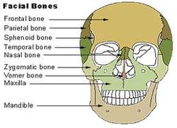
FACIAL BONES ANATOMY. The skull is additionally comprised of fourteen bones which make up the face. The facial bones do not touch the brain but are still considered part of the skull. Some cranial bones meet with the facial bones to give each individual a varying form, the frame work from which the face is then built upon.
Facial Bone Anatomy. Author: Babak Jahan-Parwar, MD; Chief Editor: Arlen D Meyers, MD, MBA more… The facial skeleton serves to protect the brain; house and protect the sense organs of smell, sight, and taste; and provide a frame on which the soft tissues of the face can act to facilitate eating, facial expression, breathing, and speech.
Overview The facial skeleton serves to protect the brain; house and protect the sense organs of smell, sight, and taste; and provide a frame on which the soft tissues of the face can act to facilitate eating, facial expression, breathing, and speech. The primary bones of the face are the mandible, maxilla, frontal bone, nasal bones, and zygoma.

Facial Bones Diagram Diagram - Facial Bones Diagram Chart - Human anatomy diagrams and charts explained. This anatomy system diagram depicts Facial Bones Diagram with parts and labels. Best diagram to help learn about health, human body and medicine.

facial bones diagram