
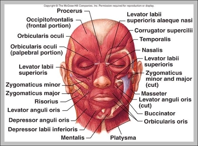
The specific location and attachments of the facial muscles enable them to produce movements of the face, such as smiling, grinning and frowning. Thus, these muscles are commonly called muscles of facial expression, or mimetic muscles. All of the facial muscles are innervated by the facial nerve (CN VII) and vascularized by the facial artery .
This field is for validation purposes and should be left unchanged. The muscles of facial expression (also known as the mimetic muscles) can generally be divided into three main functional categories: orbital, nasal and oral. These muscles are all innervated by the facial nerve (CN VII).¹
This field is for validation purposes and should be left unchanged. The muscles of facial expression (also known as the mimetic muscles) can generally be divided into three main functional categories: orbital, nasal and oral. These muscles are all innervated by the facial nerve (CN VII).¹
Muscles Of Facial Expression Image
Posted inDiagrams
Muscles Of Facial Expression Image
Post navigation
Previous Post
Next Post
Ulna Images Image 