67,060 human bone stock photos and images available, or search for human bone marrow or human bone structure to find more great stock photos and pictures. 12,251 human body organs…
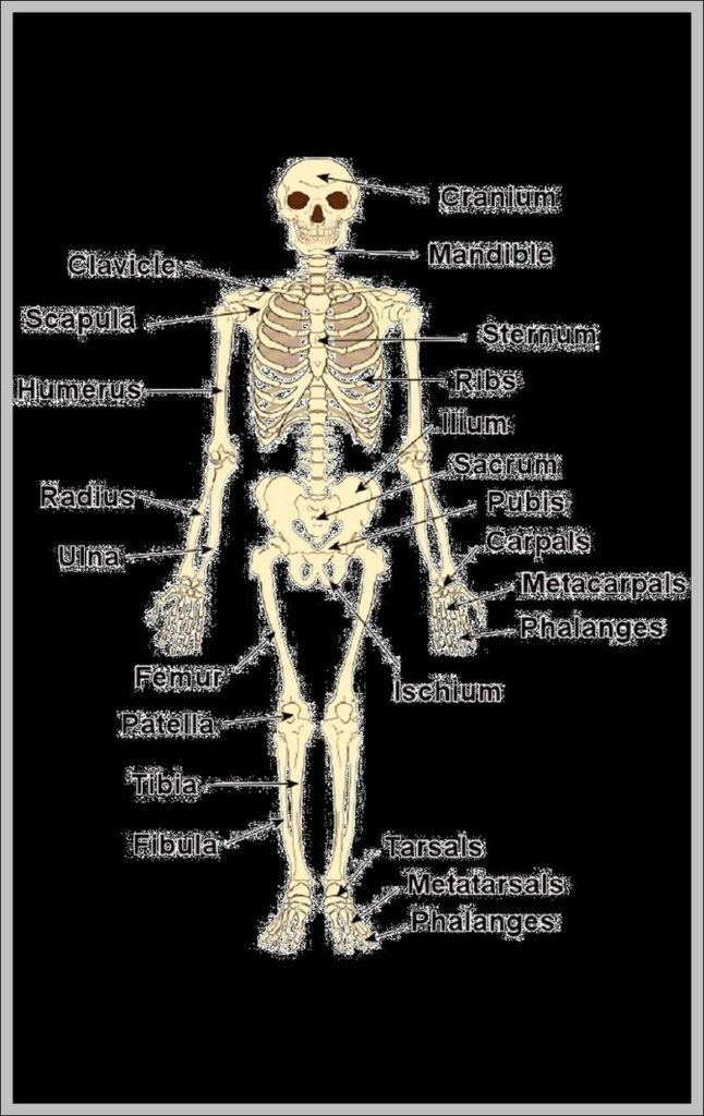
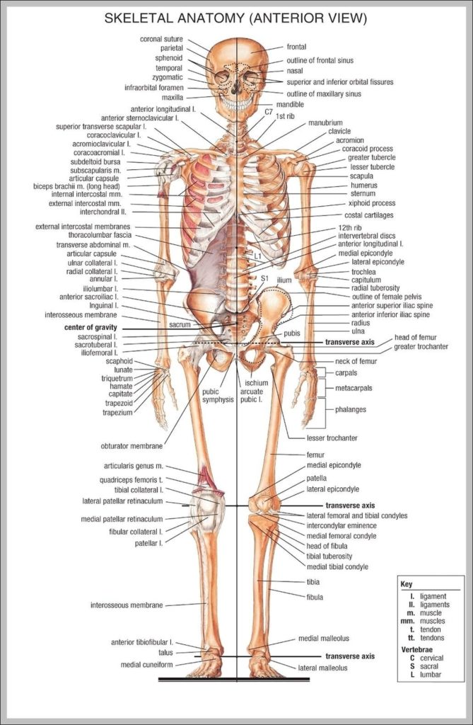
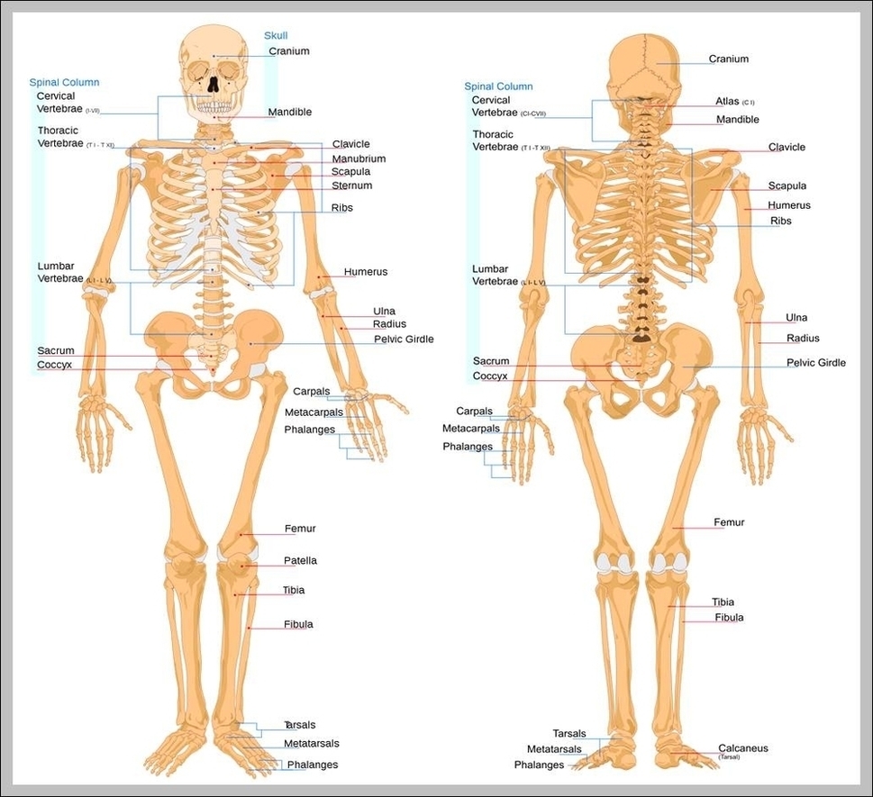
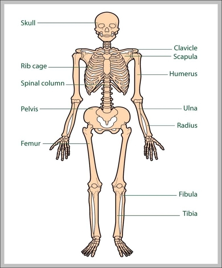
132,852 human skeleton stock photos and images available, or search for human skeleton anatomy or human skeleton vector to find more great stock photos and pictures. Male Human skeleton, four…
1,047 hip bone stock photos and images available, or search for hip bone icon or hip bone 3d to find more great stock photos and pictures. The hip is formed…
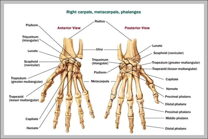
The bones of the hand and wrist provide the body with support and flexibility to manipulate objects in many different ways. Each hand contains 27 distinct bones that give the…
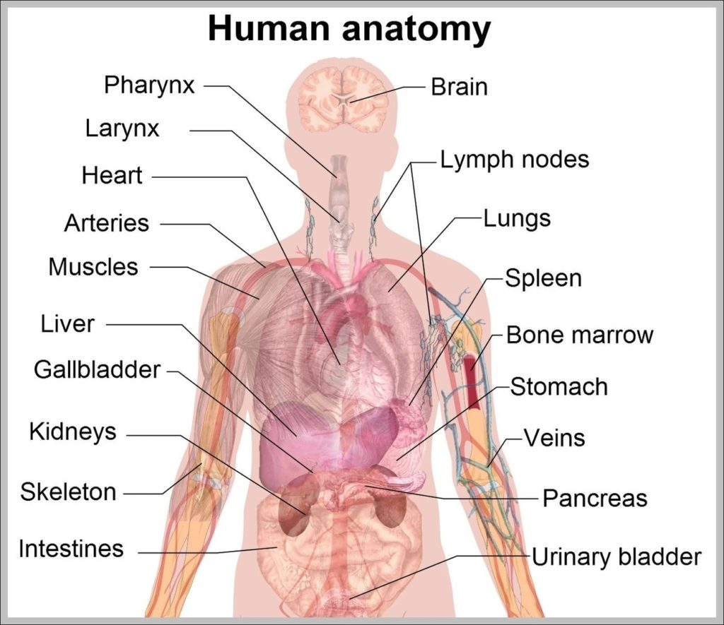
7,751 organs of the human body diagram stock illustrations and vector graphics available royalty-free, or start a new search to explore more great stock images and vector art. internal organs…
1,317 hand bone stock photos and images available, or search for skeleton hand or human bone to find more great stock photos and pictures. Carpal, metacarpal and phalanges of the…
67,060 human bone stock photos and images available, or search for human bone marrow or human bone structure to find more great stock photos and pictures. 132,852 human skeleton stock…
132,852 human skeleton stock photos and images available, or search for human skeleton anatomy or human skeleton vector to find more great stock photos and pictures. Male Human skeleton, four…
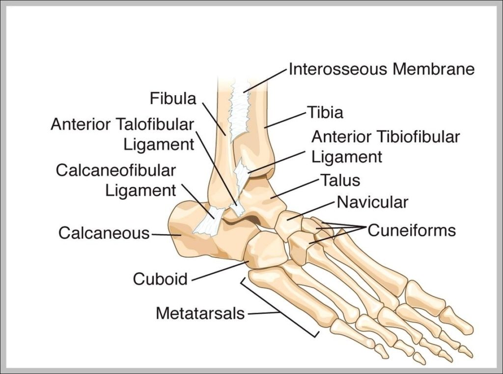
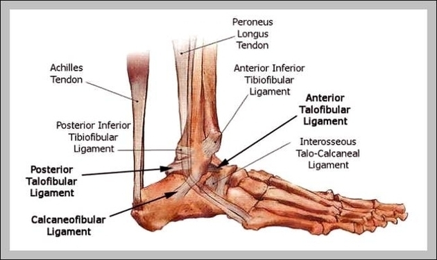
1,858 ankle bones stock photos and images available, or search for foot bones or knee to find more great stock photos and pictures. This makes the ankle one of the…
1,317 hand bone stock photos and images available, or search for skeleton hand or human bone to find more great stock photos and pictures. Carpal, metacarpal and phalanges of the…
Human lower leg muscles (soleus), illustration. The bones of the the lower leg and foot. Shown are the tibia; femur; patella; fibula, medial malleolus, lateral malleolus; metatarsals; bones; lower l…
The anatomy of the foot. The foot contains a lot of moving parts - 26 bones, 33 joints and over 100 ligaments. The foot is divided into three sections -…
67,060 human bone stock photos and images available, or search for human bone marrow or human bone structure to find more great stock photos and pictures. Abstract image human body…
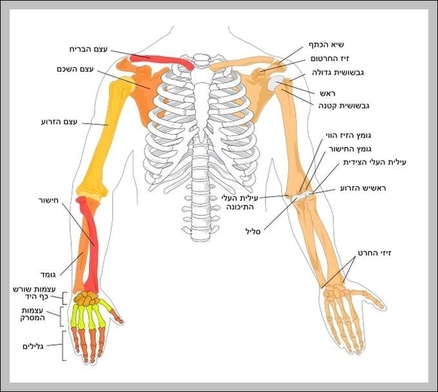
3,852 arm bone stock photos and images available or search for broken arm bone or human arm bone to find more great stock photos and pictures. Hemophilic Arthropathy Of The…
This makes the ankle one of the most stable joints in the lower extremities. Here is a brief definition of each of the ankle bones: The tibia forms the inside…
The wrist links the hand to the arm. The wrist is a complex mechanical system of 8 small bones known as the carpal bones. The carpal bones are arranged in…
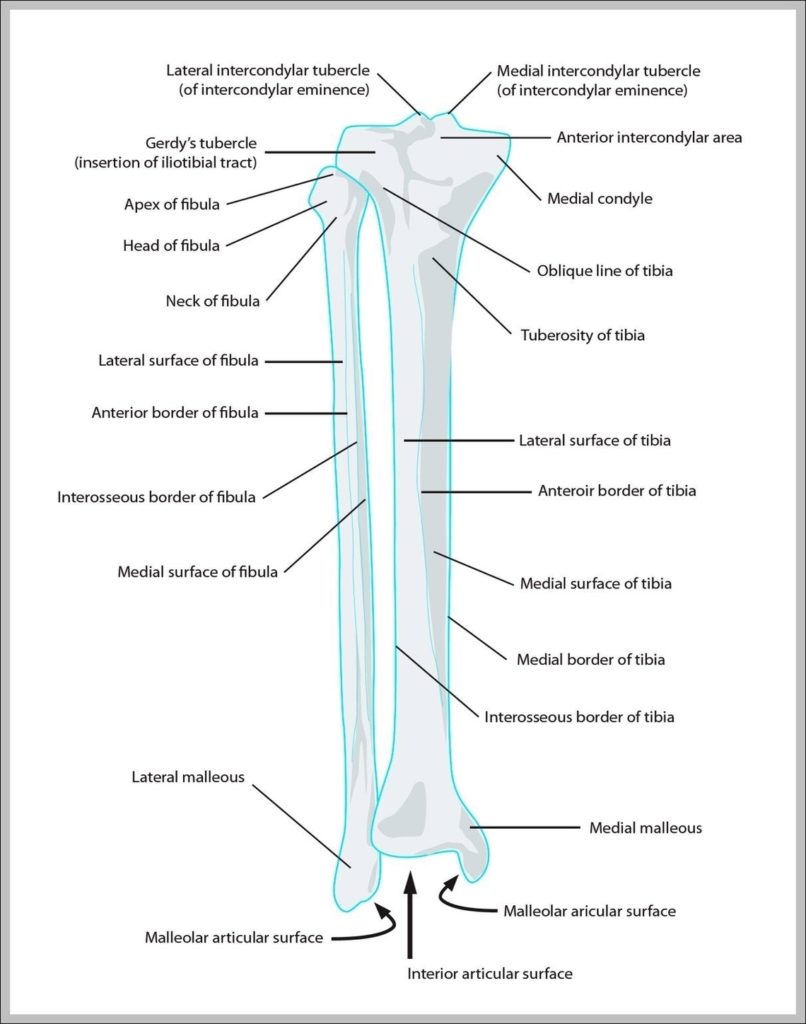
The lower leg contains two major long bones, the tibia and the fibula, which are both very strong skeletal structures. The tibia (also called the shinbone) is located near the…
It is a paired bone located at the base and lateral side of the skull. It features an important structure of the vestibulocochlear apparatus that includes the external acoustic meatus,…
Browse 2,854 human skeleton diagram stock photos and images available, or start a new search to explore more stock photos and images. human anatomy skeleton and muscles of the body…