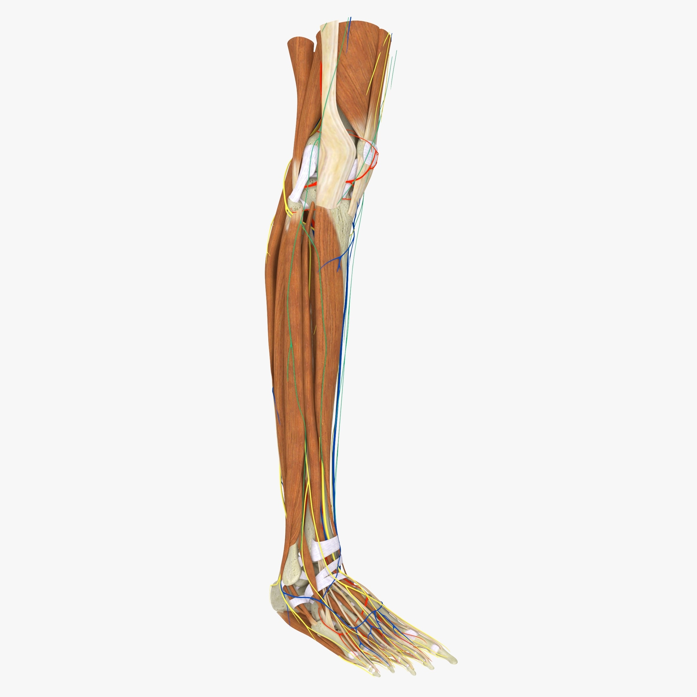
The lower leg, also known as the “leg” in anatomical terms, is a significant part of the skeletal system. It forms the lower extremity along with the upper leg and lies between the knee and the ankle.
Bones
The lower leg comprises two major long bones, the tibia and the fibula. The tibia, also known as the shinbone, is located near the midline of the leg and is the thicker and stronger of the two bones. The fibula, also referred to as the calf bone, is significantly smaller and is situated on the lateral side of the tibia.
Muscles
The main muscle in the lower leg is the gastrocnemius, which gives the calf its bulging muscular appearance. The leg muscles can be divided into anterior, lateral, and posterior groups.
Nerves
The lower leg is innervated by the superficial fibular nerve, the deep fibular nerve, and the tibial nerve, which are branches of the sacral plexus. Additionally, the common fibular (peroneal), tibial, and saphenous nerves, branches of the sciatic and femoral nerves, also serve this region.
Blood Supply
The anterior tibial, posterior tibial, and the fibular arteries supply blood to the lower leg. These blood vessels provide oxygen and nutrients to the surrounding structures, including bones, muscles, and nerves. The veins in the lower leg include the small/short saphenous, great/long saphenous, tibial, and fibular veins.
Function
The lower leg plays a crucial role in standing, walking, running, jumping, and other similar weight-bearing activities. As a result, most fractures occur in this area.
In conclusion, the lower leg is a complex structure comprising bones, muscles, nerves, and blood vessels that all work together to enable various functions. Understanding its anatomy is fundamental to appreciating its role in locomotion and weight-bearing activities.