Anatomy of the Left Hand
The human hand, a remarkable feat of engineering and evolution, is strong enough to allow climbers to tackle any mountain, yet also sufficiently precise for the manipulation of some of the world’s smallest objects and the performance of complex actions.
Bones
The hand consists of 27 distinct bones that give the hand an incredible range and precision of motion. These bones are grouped together by their location and function:
– Carpals: Eight bones that form the wrist.
– Metacarpals: Five bones that are in your palm and give it its shape.
– Phalanges: Fourteen bones that make up the segments of your fingers and thumb. Each finger has three phalanges (proximal, middle, distal), while the thumb has two (proximal, distal).
– Sesamoids: Small bones embedded in your tendons that help them move smoothly.
Muscles
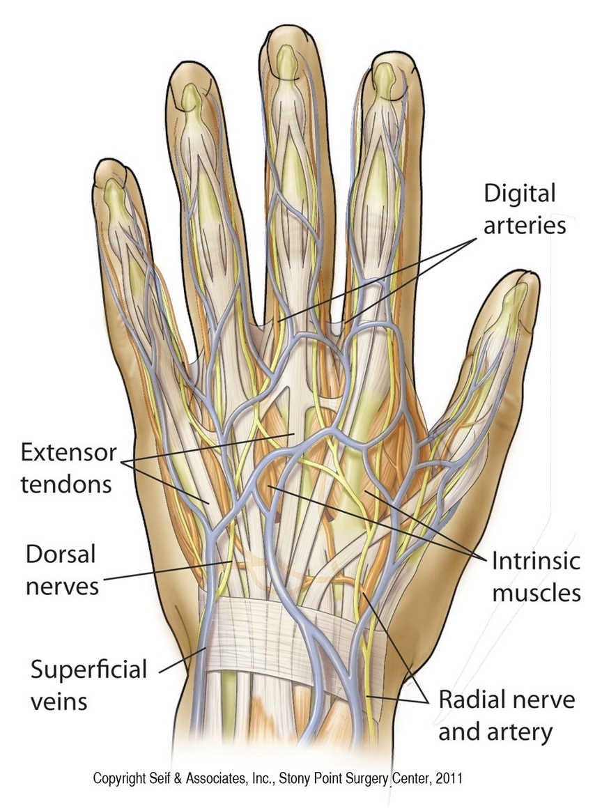
The muscles of the hand are divided into two groups: extrinsic and intrinsic. The extrinsic muscles, located in the forearm, control the gross movements of the hand and fingers. The intrinsic muscles, located within the hand itself, control the finer movements.
– Thenar group: These muscles (abductor pollicis brevis, flexor pollicis brevis, opponens pollicis) are located on the thumb side of the hand and are responsible for thumb movements.
– Hypothenar group: These muscles (abductor digiti minimi, flexor digiti minimi, opponens digiti minimi) are located on the little finger side of the hand and control the movements of the little finger.
– Metacarpal group: These muscles (lumbricals, palmar interossei, dorsal interossei) are located in the palm of the hand and control the movements of the other fingers.
Nerves
The hand is innervated by three main nerves:
– Median nerve: Predominantly supplies the thenar muscles and provides cutaneous innervation along the outside of the thumb.
– Ulnar nerve: Innervates the hypothenar and metacarpal groups. It supplies all intrinsic muscles of the hand except the LOAF muscles (Lateral two lumbricals, Opponens pollicis, Abductor pollicis brevis, Flexor pollicis brevis).
– Radial nerve: Provides sensory innervation to the back of the hand.
Blood Supply
The hand receives its blood supply from the radial and ulnar arteries, which give rise to various branches including the palmar arches (superficial, deep), palmar digital arteries (common, proper), dorsal carpal arch, dorsal metacarpal arteries, dorsal digital arteries, and the principal artery of the thumb.
Conclusion
The anatomy of the hand is complex and intricate, allowing for a wide range of movements and functions. Understanding its structure and function can provide insights into how we interact with the world around us..


