There are many types of tissues that make up joint anatomy. Typically, the bones that make up freely mobile joints are lined with articular (or hyaline) cartilage. Hyaline cartilage has a very smooth, almost frictionless surface, which allows for smooth, pain-free motion at the joint. Joints can be classified according to their degree of mobility:
Skeletal System: Bones, Joints, Cartilage, Ligaments, Bursae. The anatomy of our musculoskeletal system is quite complex. It consists of a large number of tendons, ligaments, bones, cartilage, joints, and bursae. We are able to control our muscles by sending stimulating impulses via nerves from our brain.
Skeletal System: Bones, Joints, Cartilage, Ligaments, Bursae. The anatomy of our musculoskeletal system is quite complex. It consists of a large number of tendons, ligaments, bones, cartilage, joints, and bursae. We are able to control our muscles by sending stimulating impulses via nerves from our brain.
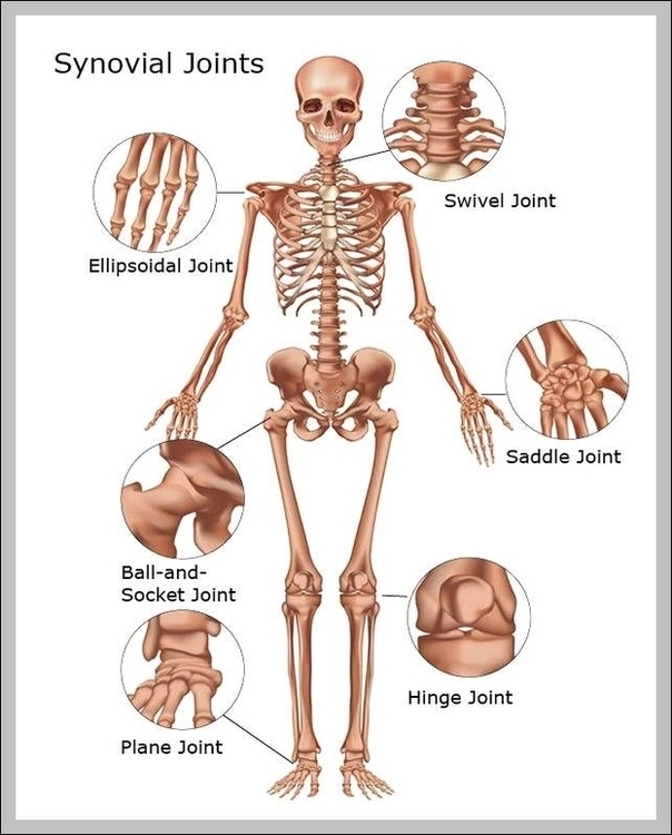
Joints In The Skeletal System Image
Posted inDiagrams
Joints In The Skeletal System Image
Post navigation
Previous Post
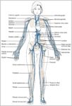 Veins In The Body Diagram Image
Next Post
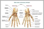
Hand Wrist Bones Image