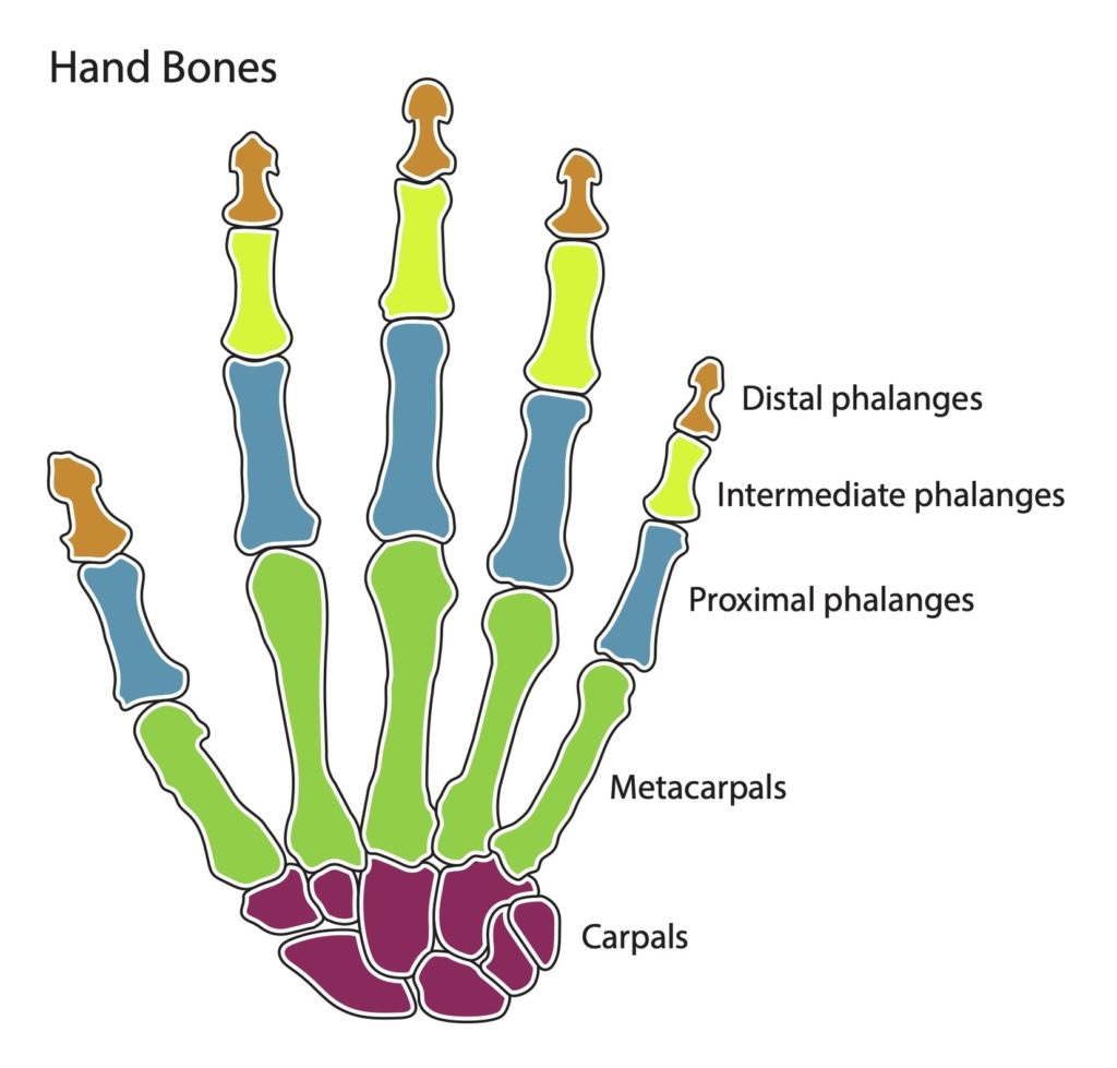
The human hand is a marvel of evolution and engineering, capable of performing a wide range of complex actions. It consists of 27 bones, divided into three categories: the carpal…
Autosomal Recessive Juvenile Parkinsonism (AR-JP) Autosomal Recessive Juvenile Parkinsonism (AR-JP), also known as PARK2 or PDJ, is a form of Parkinson's disease (PD) that was initially described in Japan. It…
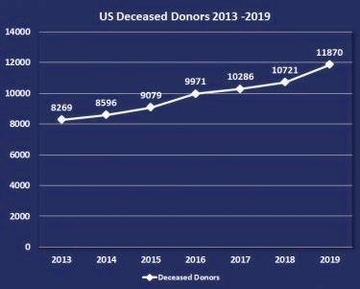
Scottish Organ Donors Organ donation in Scotland is a significant aspect of the country's healthcare system. It involves the process of giving an organ or tissue to help someone who…
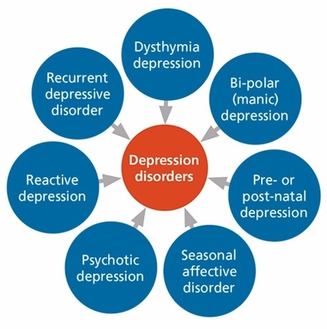
Depression is a common and serious medical illness that negatively affects how you feel, the way you think, and how you act. It causes feelings of sadness and/or a loss…
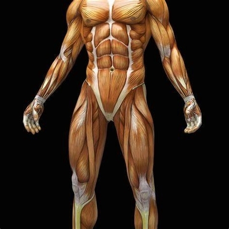
Label Muscles Worksheet A Label Muscles Worksheet is an educational tool designed to help students learn about the muscular system of the human body. It typically includes diagrams of the…
Human Body Muscles The human body is a complex system that relies on muscles for movement, posture, and balance. There are three major types of muscles in the human body:…
The lower body, also known as the lower extremity, extends from the hip to the toes and includes over 30 bones and 40 muscles. It is divided into several regions:…
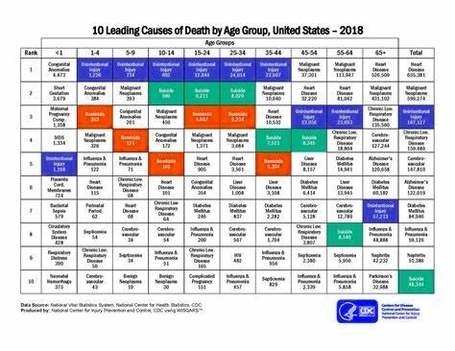
Leading Causes of Death The leading causes of death globally and in various countries are a reflection of the health challenges that populations face. These causes can be broadly categorized…
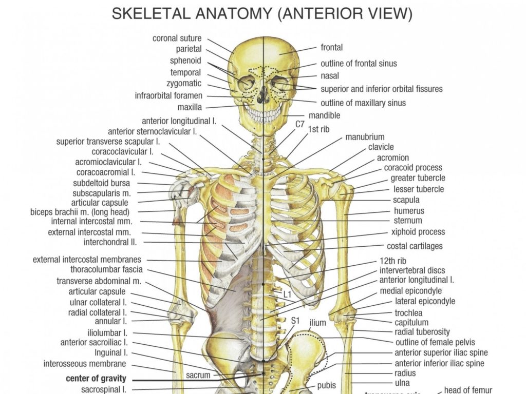
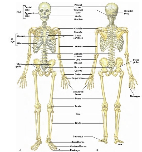
Human Bone Structure The human bone structure, also known as the skeleton, serves as the framework for the body. It consists of many individual bones and cartilages, along with bands…
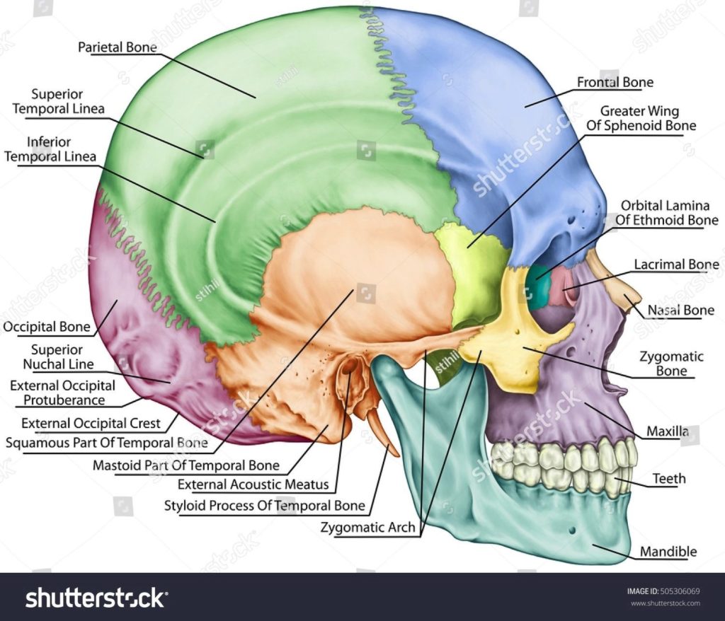
The human skull, also known as the cranium, is a complex structure that serves as the framework for the head, housing the brain and facial structures. It is composed of…
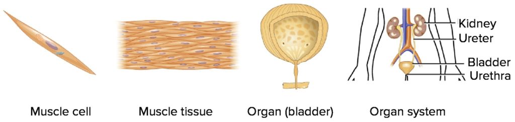
Cells, Tissues, Organs, and Systems Cells are the smallest unit of life. They come in various shapes and serve specialized purposes in the body. For instance, muscle cells contract, nerve…
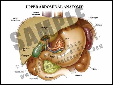
The upper abdomen, a critical region of the human body, houses several vital organs and structures. It is the area of the belly roughly between the ribs and the belly…
The human body is a complex system that is made up of different types of tissues. These tissues, which are collections of similar cells that perform a specific function, are…
Stomach Muscles and Digestive System Anatomy The stomach is a muscular organ that plays a crucial role in the digestive system. It is part of the gastrointestinal (GI) tract, which…
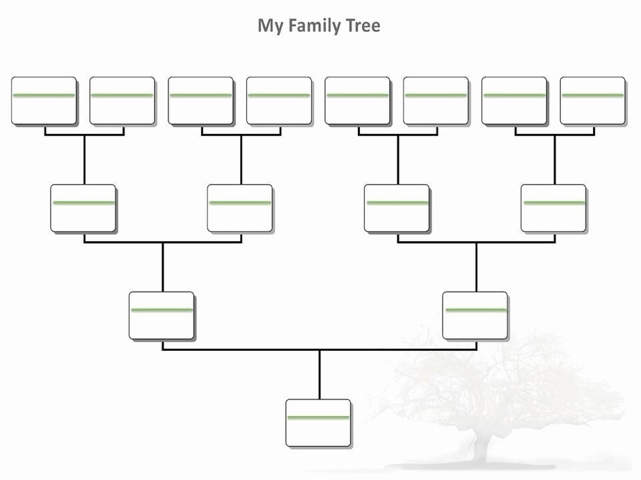
A Printable Blank Family Tree Template is a graphical representation of family relationships. It's a tool used in genealogy to visually document one's lineage, tracing relationships across several generations. These…
The microscopic structure of bone is a fascinating topic that delves into the intricate details of how bones are formed and maintained. Bone Tissue Bone tissue, also known as osseous…
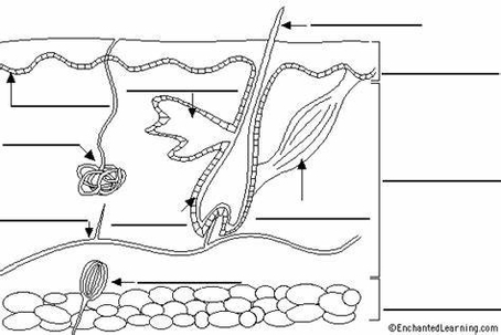
A "Label Skin Worksheet" is an educational tool designed to help students understand the structure and functions of the skin, which is a part of the integumentary system. Integumentary System…
The Human Stomach and Digestive System The human digestive system is a complex network of organs and glands that processes food. It is responsible for breaking down food into nutrients,…
Cease and Desist: An Overview A cease and desist is a formal communication sent to an individual or a company to stop a specific action and to not repeat it.…
I'm sorry, but I couldn't find any specific information related to "Low Deathdeath Row Pfp". It seems like there might be a typo or misunderstanding in your query. Could you…