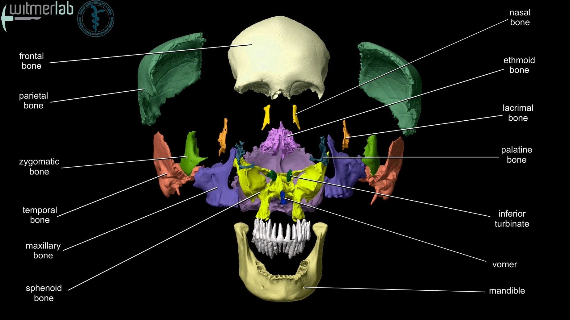
The human skull, a marvel of biological engineering, serves as the protective casing for the brain and supports the structures of the face. It consists of 22 bones, which are mostly connected by ossified joints known as sutures. The skull is divided into two main parts: the braincase (neurocranium) and the facial skeleton (viscerocranium).
Neurocranium
The neurocranium, also known as the braincase, is the upper part of the skull, which encloses and protects the brain. It consists of eight bones: the occipital, two temporal, two parietal, sphenoid, ethmoid, and frontal. The braincase is further divided into the skullcap (calvarium) and the skull base. The skullcap is made up of the pairs of parietal bones and parts of the frontal bone as well as the occipital bone.
Viscerocranium
The viscerocranium, or the facial skeleton, underlies the facial structures, forms the nasal cavity, encloses the eyeballs, and supports the teeth of the upper and lower jaws. It consists of 14 bones: the vomer, two inferior nasal conchae, two nasal, two maxillae, mandible, two palatine, two zygomatic, and two lacrimal.
utures
The most significant sutures in the human skull are the coronal suture (between the frontal and parietal bone), the sagittal suture (dividing both the parietal bones), and the lambdoidal suture (running horizontally between the occipital bone and both parietal bones).
Cranial Fossae
The skull contains three cranial fossae: anterior, middle, and posterior. The anterior cranial fossa houses the frontal lobe of the cerebral cortex, olfactory bulb, olfactory tract, and optic nerve. The middle cranial fossa contains the trochlear, abducens, oculomotor, ophthalmic, maxillary, mandibular nerves, pituitary gland, internal carotid artery, and temporal lobes of the brain. The posterior cranial fossa houses the brainstem, facial, vestibulocochlear, glossopharyngeal, vagus, accessory, hypoglossal nerves, and internal jugular vein.
Orbits
The orbits are the bony sockets that house the eyeballs and muscles that move the eyeball or open the upper eyelid. The upper margin of the anterior orbit is the supraorbital margin, which has a small opening called the supraorbital foramen for the passage of a sensory nerve to the skin of the forehead.
Nasal Cavity
Inside the nasal area of the skull, the nasal cavity is divided into halves by the nasal septum. The upper portion of the nasal septum is formed by the perpendicular plate of the ethmoid bone and the lower portion is the vomer bone.
In conclusion, the human skull is a complex structure with a primary role in protecting the brain and supporting the facial structures. Its intricate design accommodates various sensory organs and provides a framework for muscles that facilitate essential functions like eating, speaking, and expressing emotions..