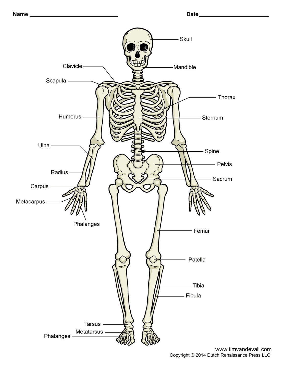
The human skeleton, an internal framework, serves as the structural support for the body. It consists of approximately 270 bones at birth, which decreases to around 206 bones by adulthood after some bones fuse together. The bone mass in the skeleton constitutes about 14% of the total body weight.
The skeleton is divided into two main subdivisions: the axial and the appendicular. The axial skeleton, which maintains the upright posture of humans, includes the vertebral column, a part of the rib cage, and the skull. The appendicular skeleton, attached to the axial skeleton, comprises the shoulder girdle, the pelvic girdle, and the bones of the upper and lower limbs.
The skeleton performs six major functions: support, movement, protection, production of blood cells, storage of minerals, and endocrine regulation. The support function is the most primitive and the oldest, with the vertebral column serving as the main support of the trunk. The joints between bones allow movement, with some allowing a wider range of movement than others.
The skeleton also provides protection. For instance, the brain is well protected by the cranium and the spinal cord by the vertebral column. The skeleton is also involved in the production of blood cells, storage of minerals, and endocrine regulation.
The human skeleton is not as sexually dimorphic as that of many other primate species, but subtle differences between sexes in the morphology of the skull, dentition, long bones, and pelvis exist. In general, female skeletal elements tend to be smaller and less robust than corresponding male elements within a given population. The human female pelvis is also different from that of males to facilitate childbirth.
In conclusion, the human skeleton is a complex and vital structure that plays a crucial role in various bodily functions. Its intricate design and functionality underscore its importance in human anatomy and physiology..