The Human Heart: An Illustrated Representation
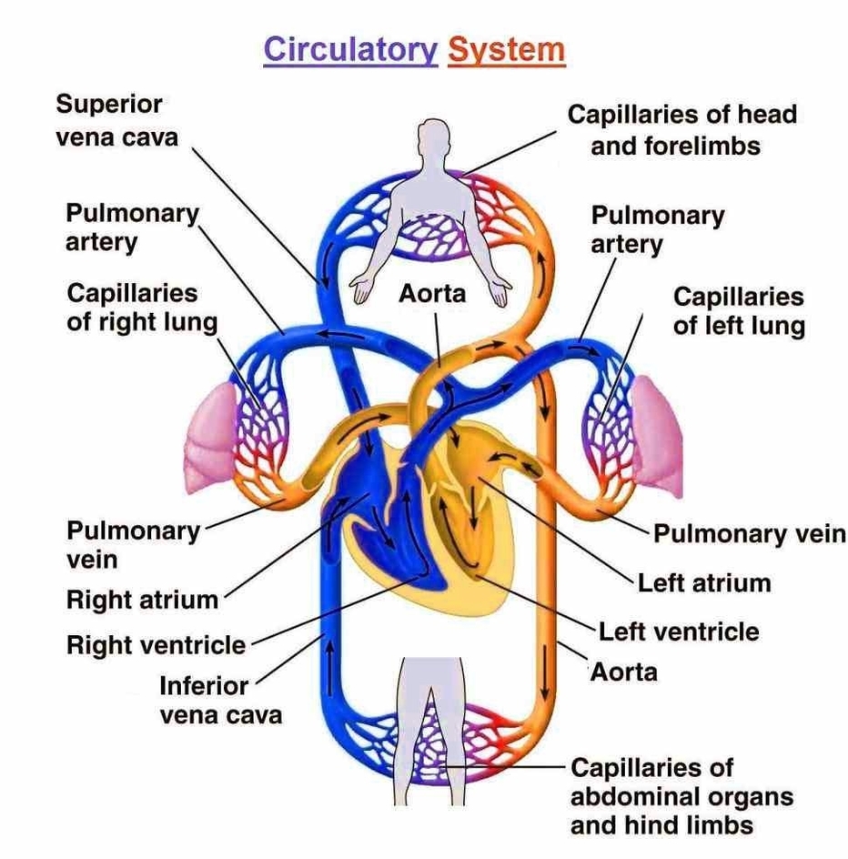
The human heart, a muscular organ roughly the size of a closed fist, serves as the body’s circulatory pump. It is located in the thoracic cavity, medial to the lungs and posterior to the sternum. The base of the heart is attached to the aorta, pulmonary arteries and veins, and the vena cava, while the apex points towards the left side.
Anatomy of the Heart
1. Pericardium: The heart resides within a fluid-filled cavity called the pericardial cavity. The pericardium, a serous membrane, lines this cavity, producing serous fluid to lubricate the heart and prevent friction.
2. Heart Wall: Comprising three layersepicardium, myocardium, and endocardiumthe heart wall encases the organ. The epicardium, also known as the visceral layer of the pericardium, forms the outermost layer.
3. Chambers and Valves: The heart consists of four chamberstwo atria (left and right) and two ventricles (left and right). The atria receive blood, while the ventricles pump blood out. Four valves (aortic, mitral, tricuspid, and pulmonary) ensure unidirectional blood flow.
4. Blood Vessels: The heart connects to several blood vessels, including the aorta, pulmonary arteries and veins, and the vena cava.
5. Conduction System: This includes the sinuatrial node, atrioventricular node, and atrioventricular bundle, which regulate the heart’s rhythm.
Illustrated Anatomy
Illustrated representations of the heart, based on medical illustrations and cadaver photography, offer an interactive way to explore heart anatomy. These illustrations, often labeled, serve as invaluable medical and anatomical tools. They depict various anatomical structures, including the myocardium, valves, coronary arteries, and the conduction system.
3D Models
3D models provide a comprehensive view of the heart’s anatomy. They allow for rotation and zooming, offering a detailed perspective of different parts, such as the aortic valve, bundle branches, chordae tendineae, and the interventricular septum.
Conclusion
Understanding the human heart’s anatomy is crucial in the medical field. Illustrated representations and 3D models serve as effective educational tools, offering an interactive and detailed exploration of the heart’s complex structure.