External Structure of the Heart
The heart, a muscular organ, pumps blood throughout the body via the circulatory system. It is located in the middle mediastinum, enclosed in a two-layered serous sac known as the pericardium. The heart’s shape resembles a quadrangular pyramid, oriented as if the pyramid has fallen onto one side. Its base faces the posterior thoracic wall, and its apex points towards the anterior thoracic wall.
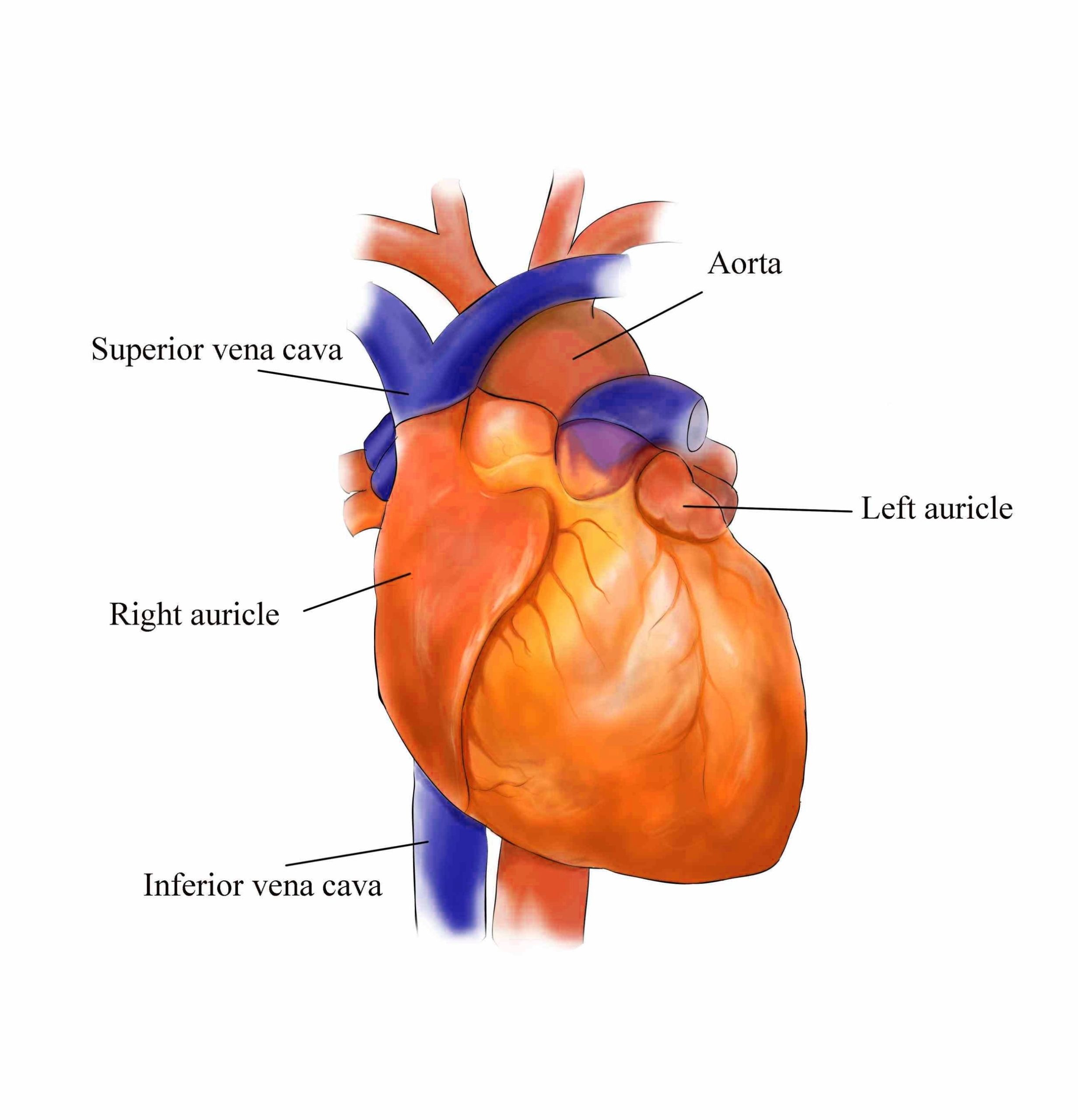
The heart has five surfaces: base (posterior), diaphragmatic (inferior), sternocostal (anterior), and left and right pulmonary surfaces. It also has several borders: right, left, superior, and inferior. The right margin is a small section of the right atrium that extends between the superior and inferior vena cava. The left margin is formed by the left ventricle and left auricle. The superior margin in the anterior view is formed by both atria and their auricles. The inferior margin is marked by the right ventricle.
The heart is divided into four chambers: two atria (right and left) and two ventricles (right and left). The right atrium and ventricle receive deoxygenated blood from systemic veins and pump it to the lungs, while the left atrium and ventricle receive oxygenated blood from the lungs and pump it to the systemic vessels, which distribute it throughout the body.
The heart’s outermost layer is the epicardium (or visceral pericardium), which covers the heart, wraps around the roots of the great blood vessels, and adheres the heart wall to a protective sac. The middle layer is the myocardium, the strong muscle tissue that powers the hearts pumping action.
The heart has four valves: tricuspid, pulmonary, mitral, and aortic. These valves ensure that blood flows in the correct direction. The heart’s blood supply comes from the right and left coronary arteries. Deoxygenated blood from the heart is drained by the coronary sinus, which includes the great, middle, and small cardiac veins, the left marginal vein, and the left posterior ventricular veins.
The heart is connected to the body’s circulatory system through several large blood vessels. The superior and inferior vena cavae carry deoxygenated blood from the body to the right atrium. The pulmonary artery carries deoxygenated blood from the right ventricle to the lungs. The pulmonary veins carry oxygenated blood from the lungs to the left atrium. The aorta carries oxygenated blood from the left ventricle to the rest of the body.
In conclusion, the heart’s external structure is complex and intricately designed to perform its vital function of pumping blood throughout the body. Its anatomy includes various surfaces, borders, chambers, valves, and blood vessels, each playing a crucial role in the heart’s operation..