External Structure of the Heart Anatomy
The heart, a muscular organ, is responsible for circulating blood throughout the body via the circulatory/vascular system. It is located in the middle mediastinum, wrapped in a two-layered serous sac known as the pericardium. The heart is shaped like a quadrangular pyramid, with its base facing the posterior thoracic wall and its apex pointed towards the anterior thoracic wall.
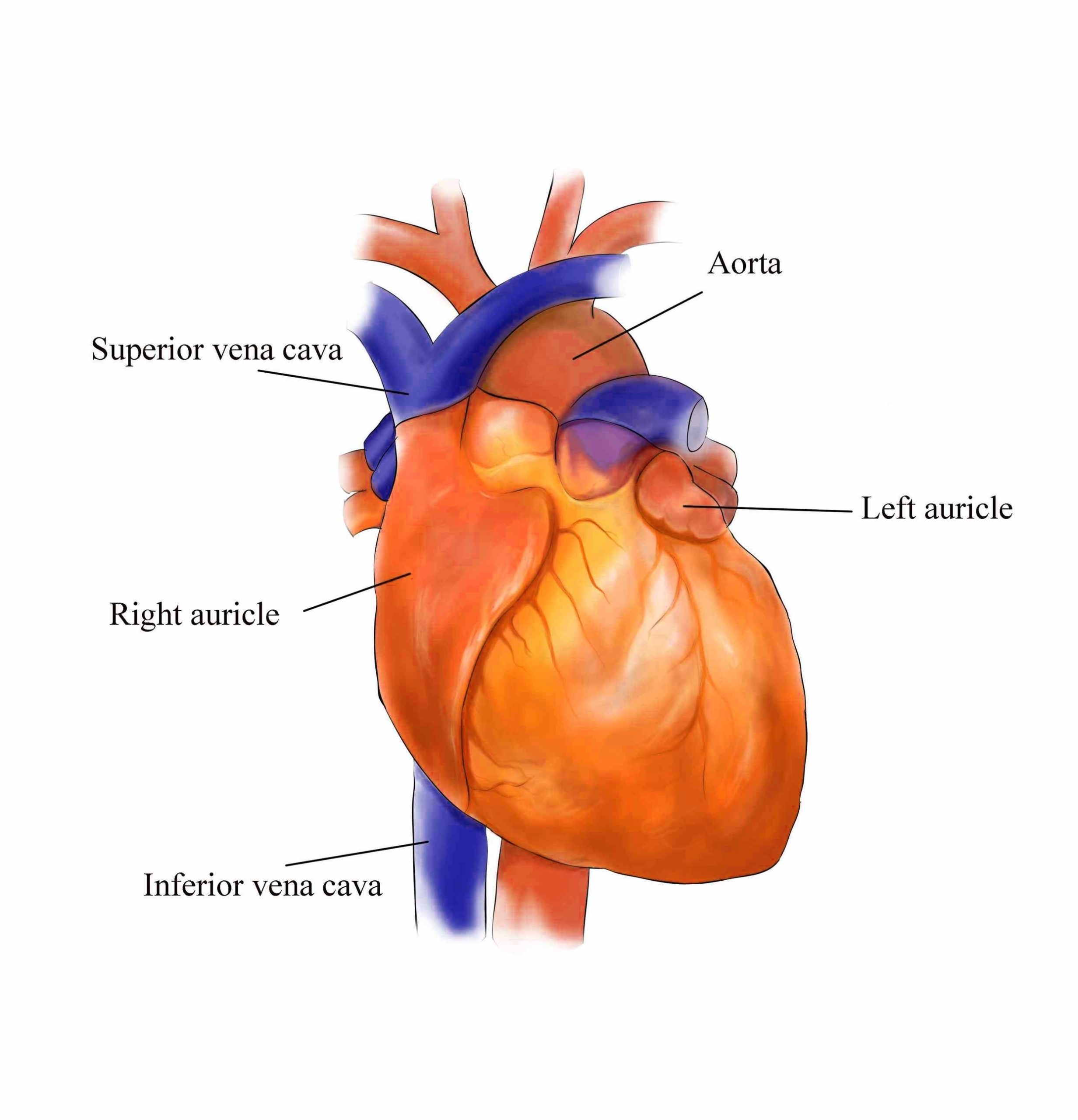
The heart’s external surface can be subdivided into five smaller surfaces that are limited by four margins. These include the base (posterior), diaphragmatic (inferior), sternocostal (anterior), and left and right pulmonary surfaces. The right margin is the small section of the right atrium that extends between the superior and inferior vena cava. The left margin is formed by the left ventricle and left auricle. The superior margin in the anterior view is formed by both atria and their auricles. The inferior margin is marked by the right ventricle.
The heart consists of several layers of a tough muscular wall, the myocardium. A thin layer of tissue, the pericardium, covers the outside, and another layer, the endocardium, lines the inside. The heart cavity is divided down the middle into a right and a left heart, which in turn are subdivided into two chambers. The upper chamber is called an atrium (or auricle), and the lower chamber is called a ventricle. The two atria act as receiving chambers for blood entering the heart; the more muscular ventricles pump the blood out of the heart.
The heart, although a single organ, can be considered as two pumps that propel blood through two different circuits. The right atrium receives venous blood from the head, chest, and arms via the large vein called the superior vena cava and receives blood from the abdomen, pelvic region, and legs via the inferior vena cava. Blood then passes through the tricuspid valve to the right ventricle, which propels it through the pulmonary artery to the lungs.
The heart’s external surfaces contain various grooves known as external sulci. The smaller upper chambers, auricles (atria) are demarcated externally from the lower larger chambers ventricles by an irregular groove called the coronary sulcus. The two ventricles are demarcated externally from each other by an oblique groove termed as inter-ventricular sulcus. This groove contains coronary blood vessels that supply blood to the heart muscles.
In conclusion, the heart’s external structure is complex and intricate, designed to efficiently perform its vital function of circulating blood throughout the body. Its unique shape, surfaces, and margins, along with the various chambers and valves, all contribute to its remarkable functionality..