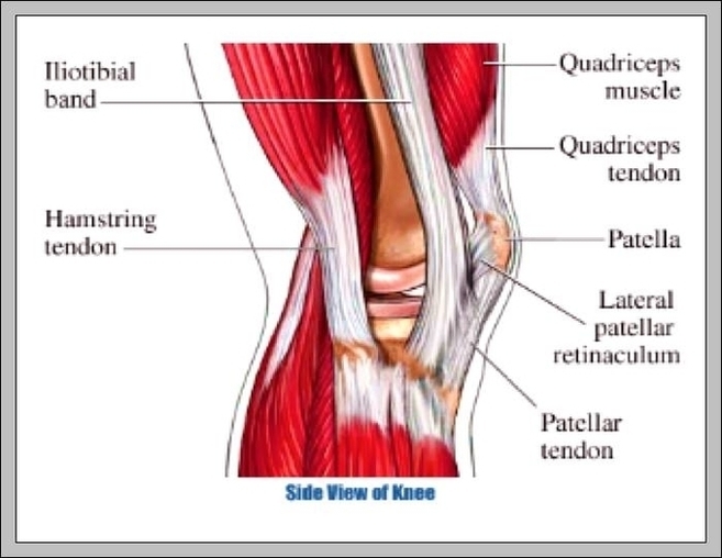
To understand one of the most complex joints of our body i.e. the knee joint, you need a perfectly labeled diagram of the knee. This will help you to understand the mechanism as well as the working.
Knee joint is one of the most important hinge joints of our body. Its complexity and its efficiency is the best example of God’s creation. The anatomy of the knee consists of bones, muscles, nerves, cartilages, tendons and ligaments. All these parts combine and work together.
The hamstrings and quadriceps work together, one contracting (agonist) while the other relaxes (antagonist) to allow the knee to bend and straighten. Here we look at each of the muscles of the knee, how they work, what can go wrong and how to prevent knee muscle injuries.

Diagram Of The Knee Muscles Image
Posted inDiagrams


