The Human Eye (Eyeball) Diagram, Parts and Pictures. The human eye consists of the eyeball, optic nerve, orbit and appendages (eyelids, extraocular muscles and lacrimal glands). While the eyeball is the actual sensory organ, the other parts of of the eye are equally important in maintaining the health and function of the eye as a whole.
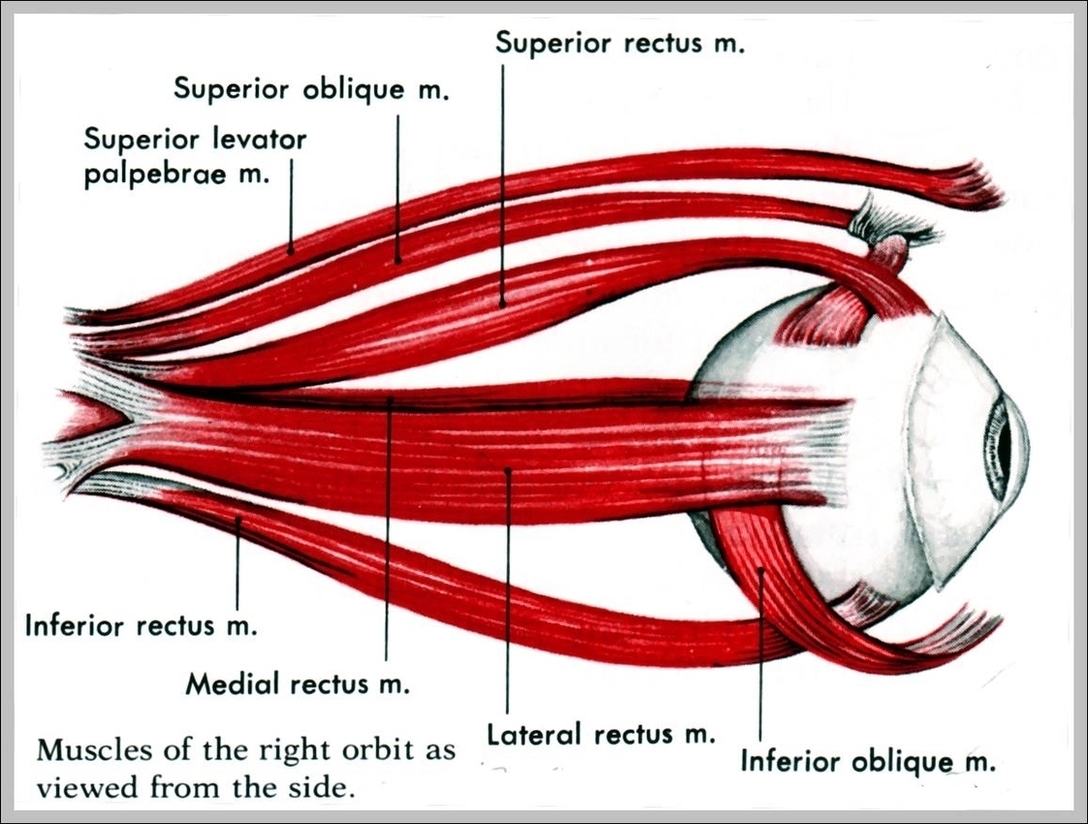
There are two types of eye muscles: extrinsic muscles that control eye movement and position, and intrinsic muscles that control near focusing and how much light enters the eye. Extrinsic eye muscles (also called extraocular muscles) are attached to the outside of the eyeball and enable the eyes to move in all directions of sight.
There are two types of eye muscles: extrinsic muscles that control eye movement and position, and intrinsic muscles that control near focusing and how much light enters the eye. Extrinsic eye muscles (also called extraocular muscles) are attached to the outside of the eyeball and enable the eyes to move in all directions of sight.

Diagram Of Eye Muscles Image
Posted inDiagrams

