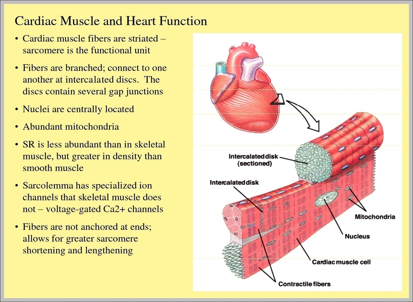
Cardiac muscle tissue is only found in your heart, where it performs coordinated contractions that allow your heart to pump blood through your circulatory system. Keep reading to learn more about the function and structure of cardiac muscle tissue, as well as conditions that affect this type of muscle tissue. How does it function?
Cardiac Muscle Definition Cardiac muscle, also known as heart muscle, is the layer of muscle tissue which lies between the endocardium and epicardium. These inner and outer layers of the heart, respectively, surround the cardiac muscle tissue and separate it from the blood and other organs.
Cardiac Muscle Definition Cardiac muscle, also known as heart muscle, is the layer of muscle tissue which lies between the endocardium and epicardium. These inner and outer layers of the heart, respectively, surround the cardiac muscle tissue and separate it from the blood and other organs.

Cardiac Tissue Function Image
Posted inDiagrams


