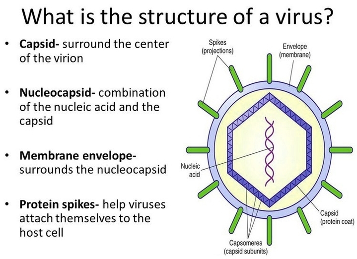
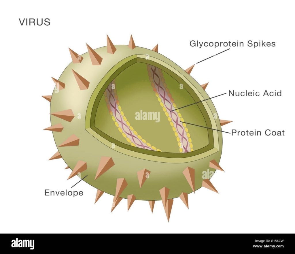
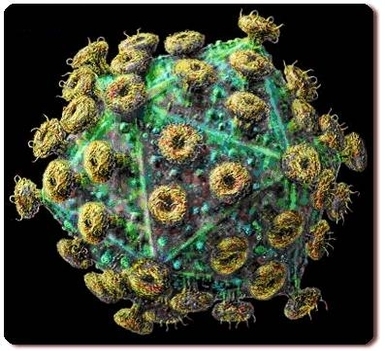
Structure of a Virus A virus is a microscopic infectious agent that replicates only inside the living cells of an organism. Viruses can infect all types of life forms, from…
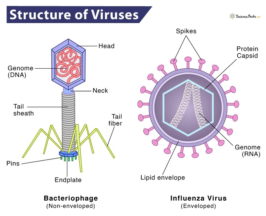
Structure of Viruses Viruses are tiny, infectious particles that can reproduce only by infecting a host cell. They are much smaller than bacteria and consist of a single- or double-stranded…
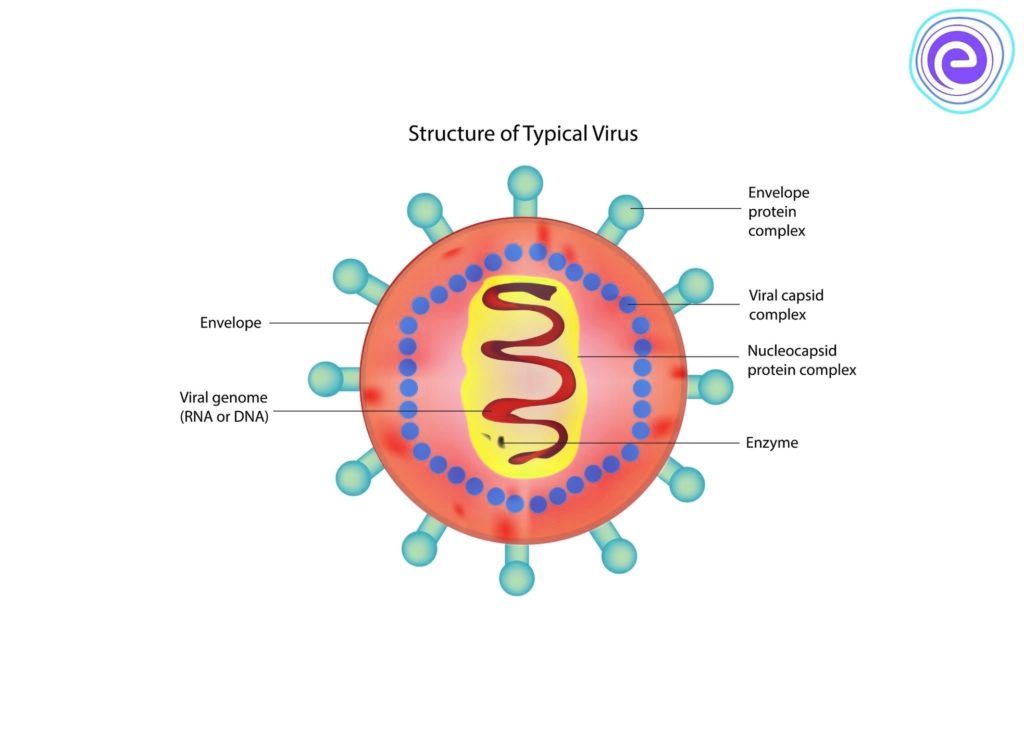
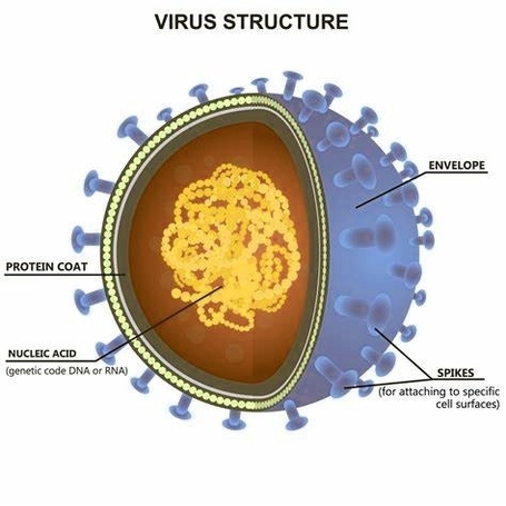
Internal Structure of a Virus A virus is a microscopic infectious particle that can reproduce only by infecting a host cell. Viruses are not considered living as they can't reproduce…
Virus Cell Structure Viruses are unique entities that straddle the line between living and non-living. They are much smaller than cells and are composed of a nucleic acid genome (either…
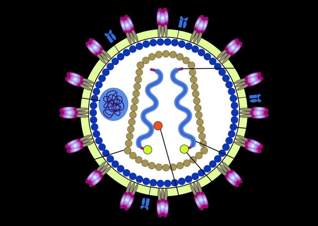
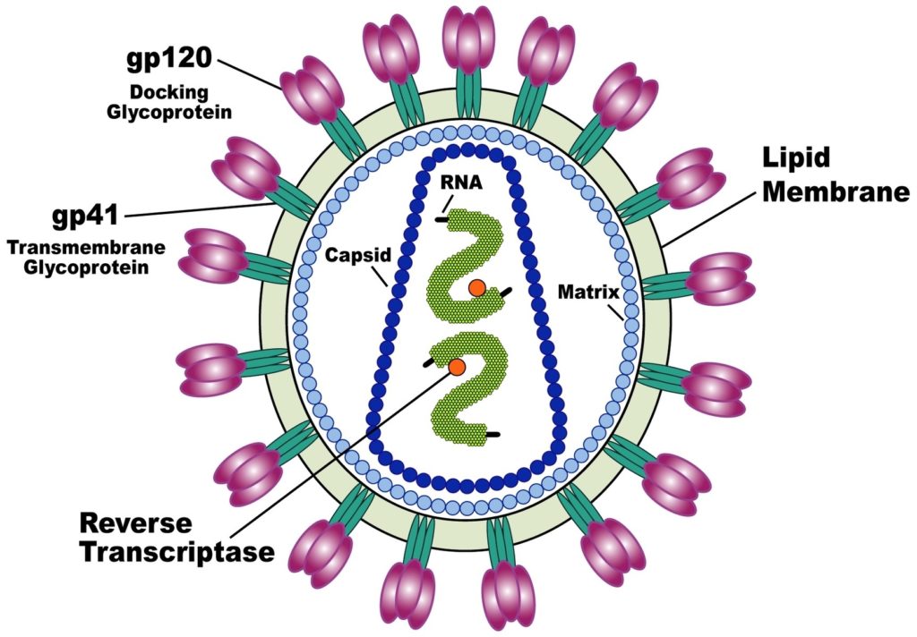
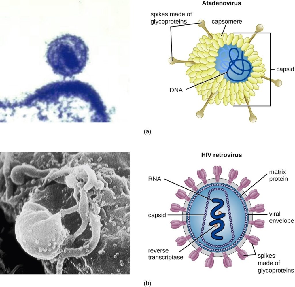
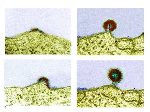
HIV Virus Structure The Human Immunodeficiency Virus (HIV) is a complex retrovirus with a unique structure that plays a crucial role in its ability to infect human cells and cause…
HIV Virus Structure The Human Immunodeficiency Virus (HIV) is a complex retrovirus that has been extensively studied since its discovery in 1983. The structure of HIV is unique and different…
The Structure of the HIV Virus The Human Immunodeficiency Virus (HIV) is a complex entity that has been the subject of extensive research since its discovery in 1983. The structure…
Virus Cell Structure A virus is a tiny, infectious particle that can reproduce only by infecting a host cell. Viruses are much smaller than bacteria and consist of a single-…
There are three stages of HIV infection: Acute HIV Infection Acute HIV infectionis the earliest stage of HIV infection, and it generally develops within 2 to 4 weeks after infection…
HIV is a virus spread through certain body fluids that attacks the body’s immune system, specifically the CD4 cells, often called T cells. Over time, HIV can destroy so many…