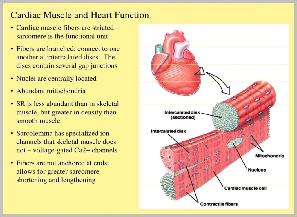
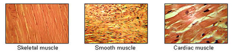
Kidney Tissue Image

98 muscular system labeled diagram stock photos and images available, or start a new search to explore more stock photos and images. Frontal view of the muscular system of the male human body with descriptive labels pointing to the muscles on a white background. Labeled Anatomy Chart of Neck and Shoulder Muscles on White…
Muscular System Labeling Worksheet Somatic signals are sent from the cerebral cortex to nerves associated with specific skeletal muscles. This results in muscular hypertrophy which is a result in a increase of myofibrils as a result of increased exercise level of physical activity determined by the frequency of recruit ment and the load.

Kidney Tissue Image Diagram - Kidney Tissue Image Chart - Human anatomy diagrams and charts explained. This anatomy system diagram depicts Kidney Tissue Image with parts and labels. Best diagram to help learn about health, human body and medicine.

Kidney Tissue Image

Cardiac Tissue Function Image

Cardiac muscle tissue is only found in your heart, where it performs coordinated contractions that allow your heart to pump blood through your circulatory system. Keep reading to learn more about the function and structure of cardiac muscle tissue, as well as conditions that affect this type of muscle tissue. How does it function?
Cardiac Muscle Definition Cardiac muscle, also known as heart muscle, is the layer of muscle tissue which lies between the endocardium and epicardium. These inner and outer layers of the heart, respectively, surround the cardiac muscle tissue and separate it from the blood and other organs.

Cardiac Tissue Function Image Diagram - Cardiac Tissue Function Image Chart - Human anatomy diagrams and charts explained. This anatomy system diagram depicts Cardiac Tissue Function Image with parts and labels. Best diagram to help learn about health, human body and medicine.

Cardiac Tissue Function Image

3 Types Of Muscle Tissue Image

3 Types of Muscle Tissue: The Function of Skeletal, Cardiac, and Smooth Muscle. About half of your body’s weight is muscle. In the muscular system, muscle tissue is categorized into three distinct types: skeletal, cardiac, and smooth. Each type of muscle tissue in the human body has a unique structure and a specific role.
Skeletal muscle is striated muscle. Unlike cardiac muscle, the cells are not branched. Skeletal muscle cells are covered by connective tissue, which protects and supports muscle fiber bundles.

3 Types Of Muscle Tissue Image Diagram - 3 Types Of Muscle Tissue Image Chart - Human anatomy diagrams and charts explained. This anatomy system diagram depicts 3 Types Of Muscle Tissue Image with parts and labels. Best diagram to help learn about health, human body and medicine.

3 Types Of Muscle Tissue Image

Epithelium Tissue Diagram

Structure of Epithelial Tissue Epithelial tissue is formed from a tightly fitted continuous layer of cells. One surface of the epithelial tissue is exposed to either the external environment or the body fluid. The other surface is attached to tissue by a membrane, which consists of fibres and polysaccharides secreted by epithelial cells.
Simple Columnar Epithelium: A Labeled Diagram and Functions Epithelium is a tissue that lines the internal surface of the body, as well as the internal organs. Simple epithelium is one of the types of epithelium that is divided into simple columnar epithelium, simple squamous epithelium, and simple cuboidal epithelium.
It supports the overlying epithelia, provides a surface along which epithelial cells migrate across during growth and wound healing, and it acts as a physical barrier (to malignant melanoma) Secreted by the epithelial cells.

Epithelium Tissue Diagram Diagram - Epithelium Tissue Diagram Chart - Human anatomy diagrams and charts explained. This anatomy system diagram depicts Epithelium Tissue Diagram with parts and labels. Best diagram to help learn about health, human body and medicine.

epithelium tissue diagram

Muscle Tissue Diagram

Updated December 8, 2017. Muscle tissue is a specialized tissue found in animals which functions by contracting, thereby applying forces to different parts of the body. Muscle tissue consists of fibers of muscle cells connected together in sheets and fibers.
Skeletal muscle tissue is arranged in bundles surrounded by connective tissue. Under the light microscope, muscle cells appear striated with many nuclei squeezed along the membranes.
The muscle cell, or myocyte, develops from myoblasts derived from the mesoderm. Myocytes and their numbers remain relatively constant throughout life. Skeletal muscle tissue is arranged in bundles surrounded by connective tissue. Under the light microscope, muscle cells appear striated with many nuclei squeezed along the membranes.

Muscle Tissue Diagram Diagram - Muscle Tissue Diagram Chart - Human anatomy diagrams and charts explained. This anatomy system diagram depicts Muscle Tissue Diagram with parts and labels. Best diagram to help learn about health, human body and medicine.

muscle tissue diagram

Bone Tissue Diagram

Structure of Bone Tissue. There are two types of bone tissue: compact and spongy. The names imply that the two types differ in density, or how tightly the tissue is packed together. There are three types of cells that contribute to bone homeostasis.
Figure 6.3.3 – Anatomy of a Flat Bone: This cross-section of a flat bone shows the spongy bone (diploë) covered on either side by a layer of compact bone. Osseous tissue is a connective tissue and like all connective tissues contains relatively few cells and large amounts of extracellular matrix.
Four types of cells are found within bone tissue. Osteogenic cells are undifferentiated and develop into osteoblasts. When osteoblasts get trapped within the calcified matrix, their structure and function changes, and they become osteocytes.

Bone Tissue Diagram Diagram - Bone Tissue Diagram Chart - Human anatomy diagrams and charts explained. This anatomy system diagram depicts Bone Tissue Diagram with parts and labels. Best diagram to help learn about health, human body and medicine.

bone tissue diagram