Diagram Of Thoracic Vertebrae Image

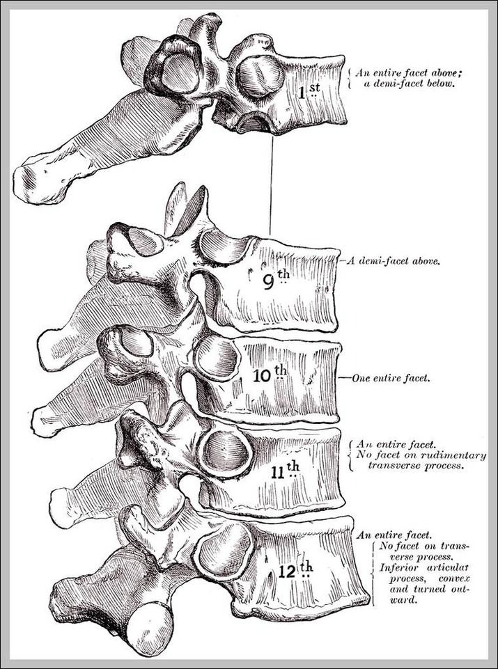
This article will elucidate all the mysteries surrounding the thoracic vertebrae and will describe both their typical and atypical features. The thoracic vertebrae are located in the middle section of the vertebral column, specifically inferior to the cervical vertebrae and superior to the lumbar vertebrae.
368 vertebrae diagram stock photos and images available, or start a new search to explore more stock photos and images. An anatomical diagram showing the human vertebral column, viewed from the left side, circa 1930.

Diagram Of Thoracic Vertebrae Image Diagram - Diagram Of Thoracic Vertebrae Image Chart - Human anatomy diagrams and charts explained. This anatomy system diagram depicts Diagram Of Thoracic Vertebrae Image with parts and labels. Best diagram to help learn about health, human body and medicine.

Diagram Of Thoracic Vertebrae Image

Thoracic Vertebra Image

This article will elucidate all the mysteries surrounding the thoracic vertebrae and will describe both their typical and atypical features. The thoracic vertebrae are located in the middle section of the vertebral column, specifically inferior to the cervical vertebrae and superior to the lumbar vertebrae.
T1 (1st Thoracic Vertebra) The T1 vertebra is the first (uppermost) of the twelve (12) thoracic vertebrae that make up the central and largest section of the spinal column between the lumbar vertebrae below and the cervical vertebrae above. While larger than the C7 vertebra above it, the T1 is the smallest of the thoracic vertebrae.

Thoracic Vertebra Image Diagram - Thoracic Vertebra Image Chart - Human anatomy diagrams and charts explained. This anatomy system diagram depicts Thoracic Vertebra Image with parts and labels. Best diagram to help learn about health, human body and medicine.

Thoracic Vertebra Image

Thoracic Vertebrae Numbers Image

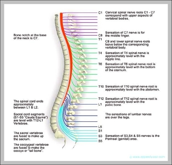
The thoracic vertebrae are bones located between the cervical and lumbar vertebrae. There are 12 thoracic vertebrae in humans, and these bones increase in size as you move down the body.
9,447 thoracic spine stock photos, vectors, and illustrations are available royalty-free. See thoracic spine stock video clips

Thoracic Vertebrae Numbers Image Diagram - Thoracic Vertebrae Numbers Image Chart - Human anatomy diagrams and charts explained. This anatomy system diagram depicts Thoracic Vertebrae Numbers Image with parts and labels. Best diagram to help learn about health, human body and medicine.

Thoracic Vertebrae Numbers Image

Thoracic Duct Function Image

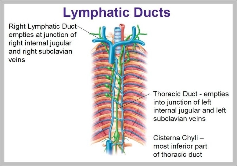
The thoracic duct carries chyle, a liquid containing both lymph and emulsified fats, rather than pure lymph. It also collects most of the lymph in the body other than from the right thorax, arm, head, and neck (which are drained by the right lymphatic duct ).
Variant anatomy. The thoracic duct has variant anatomy in ~40% (range 30-50%) of the population 1, 4: double thoracic ducts (i.e. multiple ducts) (~20%) 1, 2, 4 aberrant termination: left internal jugular vein, left external jugular vein, azygos vein, brachiocephalic vein or left subclavian vein 1, 4.

Thoracic Duct Function Image Diagram - Thoracic Duct Function Image Chart - Human anatomy diagrams and charts explained. This anatomy system diagram depicts Thoracic Duct Function Image with parts and labels. Best diagram to help learn about health, human body and medicine.

Thoracic Duct Function Image

Thoracic Spine Anatomy Pictures Image

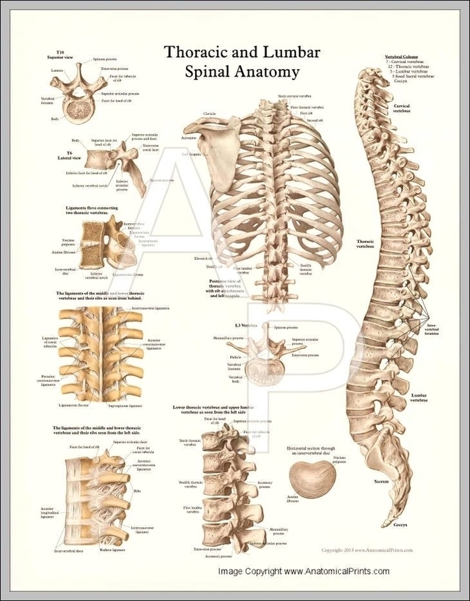
Anatomy of the Thoracic Spine. In technical terms, your spinal column at the mid and upper back levels is called the thoracic spine. The thoracic spine is comprised of 12 spinal bones connected to and occupying the same level in your body as your 12 ribs.
9,447 thoracic spine stock photos, vectors, and illustrations are available royalty-free. See thoracic spine stock video clips

Thoracic Spine Anatomy Pictures Image Diagram - Thoracic Spine Anatomy Pictures Image Chart - Human anatomy diagrams and charts explained. This anatomy system diagram depicts Thoracic Spine Anatomy Pictures Image with parts and labels. Best diagram to help learn about health, human body and medicine.

Thoracic Spine Anatomy Pictures Image

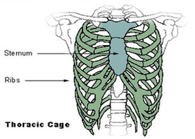
Thoracic Cage Diagram

The angles of the ribs form the most posterior extent of the thoracic cage. In the anatomical position, the angles align with the medial border of the scapula. A shallow costal groove for the passage of blood vessels and a nerve is found along the inferior margin of each rib. The bony ribs do not extend anteriorly completely around to the sternum.
The thoracic cage consists of ribs and the sternum. It is a bony structure that protects the organs in the thoracic cavity. It includes 25 bones, i.e., 1 sternum and 24 ribs. A. Ribs: They are a cage-like formation of thin, flat and curved bones.
The potential for movement is related to the flexibility provided by the ribs and their joints. The thoracic cage is composed of the thoracic skeleton, which includes the sternum, 12 pairs of ribs and 12 thoracic vertebrae, associated with the costal cartilages and intervertebral discs, respectively.

Thoracic Cage Diagram Diagram - Thoracic Cage Diagram Chart - Human anatomy diagrams and charts explained. This anatomy system diagram depicts Thoracic Cage Diagram with parts and labels. Best diagram to help learn about health, human body and medicine.

thoracic cage diagram