Human lower leg muscles (soleus), illustration. The bones of the the lower leg and foot. Shown are the tibia; femur; patella; fibula, medial malleolus, lateral malleolus; metatarsals; bones; lower l…
Your lower back (lumbar spine) is the anatomic region between your lowest rib and the upper part of the buttock. 1 Your spine in this region has a natural inward…
WebMD's Intestines Anatomy Page provides a detailed image and definition of the intestines. Learn about its parts, location in the body, function, and conditions that affect the intestines. home image…
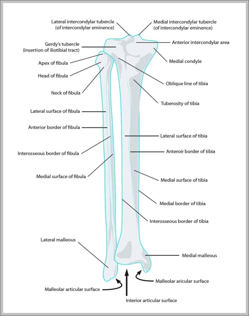
The lower leg contains two major long bones, the tibia and the fibula, which are both very strong skeletal structures. The tibia (also called the shinbone) is located near the…
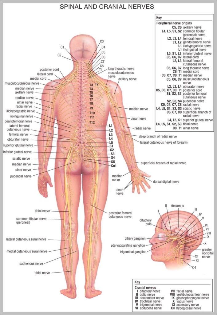
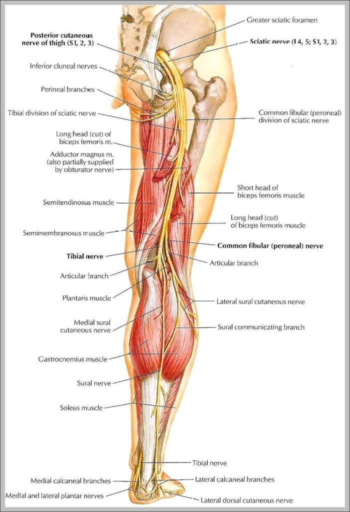
There are two main nerves in the leg: the femoral nerve serves the front and the sciatic nerve controls the back of the leg. The nerves of the leg can…