Leg Muscles And Ligaments

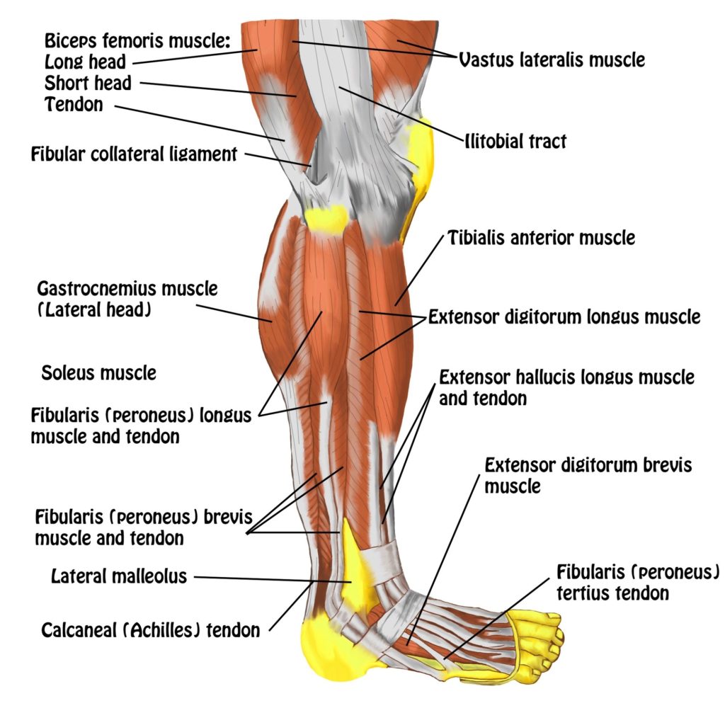
Leg Muscles and Ligaments

The leg, anatomically defined as the region of the lower limb below the knee, is a complex structure that includes various muscles and ligaments. These components work together to enable movements such as walking, running, jumping, and flexing the feet.

Muscles of the Leg

The leg muscles are organized into three groups:

1. Anterior (dorsiflexor) group: This includes the tibialis anterior, extensor digitorum longus, fibularis tertius, and extensor hallucis longus. These muscles help lift and lower your foot, extend your toes, and are crucial for the swing phase of the gait cycle.

2. Posterior (plantar flexor) group: Comprising of the gastrocnemius, plantaris, soleus, tibialis posterior, flexor hallucis longus, popliteus, and flexor digitorum longus. These muscles help flex and point your toes, jump, run, push off into a sprint, lock and unlock your knee, maintain good posture by stabilizing your legs, and stand up straight by supporting the arch of your foot.

3. Lateral (fibular) group: Consists of the fibularis longus and fibularis brevis. These muscles stabilize your foot when walking or running and allow you to move your foot from side to side.

Ligaments of the Leg

Ligaments are connective tissues that help support and stabilize joints. They are crucial for the proper functioning of the leg, providing stability and facilitating movement.

Conclusion

The leg’s muscles and ligaments play a vital role in our daily activities, from simple tasks like standing and walking to more complex actions like running and jumping. They work in harmony to provide stability, support body weight, and enable movement. Understanding their structure and function is essential for maintaining leg health and managing potential injuries..

Leg Muscles And Ligaments Diagram - Leg Muscles And Ligaments Chart - Human anatomy diagrams and charts explained. This anatomy system diagram depicts Leg Muscles And Ligaments with parts and labels. Best diagram to help learn about health, human body and medicine.

Leg Muscles And Ligaments

Ligaments Of Elbow Image

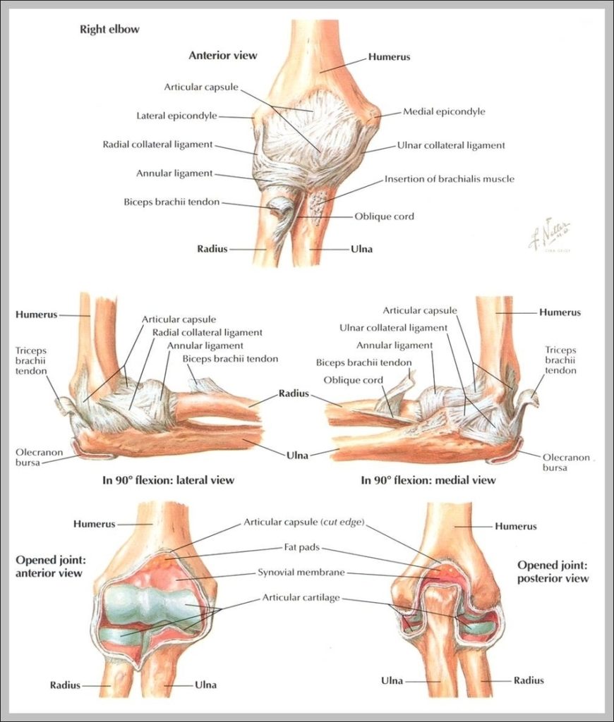
Ulnar collateral ligament or UCL, lateral collateral ligament and annular ligament form the ligaments in elbow. Here we will look in detail about the ligaments, the common injuries affecting them, how they are diagnosed and treated.
The annular ligament of the elbow extends from the ulna around the head of the radius to hold the bones of the lower arm together. These ligaments allow for movement and stretching of the elbow while resisting dislocation of the bones.

Ligaments Of Elbow Image Diagram - Ligaments Of Elbow Image Chart - Human anatomy diagrams and charts explained. This anatomy system diagram depicts Ligaments Of Elbow Image with parts and labels. Best diagram to help learn about health, human body and medicine.

Ligaments Of Elbow Image

Hand Ligaments And Tendons Image

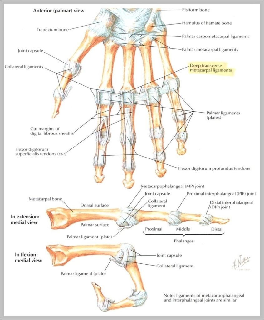
Ligaments. There are many ligament of the hand that are made up of tough bands of fibrous tissue. As there are many small bones and joints in the hand, there are also many ligaments within the hand that help to keep the bones together and stabilize the hand.
324 hand anatomy tendons stock photos and images available, or start a new search to explore more stock photos and images. Vintage anatomical color illustration of the musculature of the human forearm.

Hand Ligaments And Tendons Image Diagram - Hand Ligaments And Tendons Image Chart - Human anatomy diagrams and charts explained. This anatomy system diagram depicts Hand Ligaments And Tendons Image with parts and labels. Best diagram to help learn about health, human body and medicine.

Hand Ligaments And Tendons Image

Ligaments Of The Shoulder Joint Image

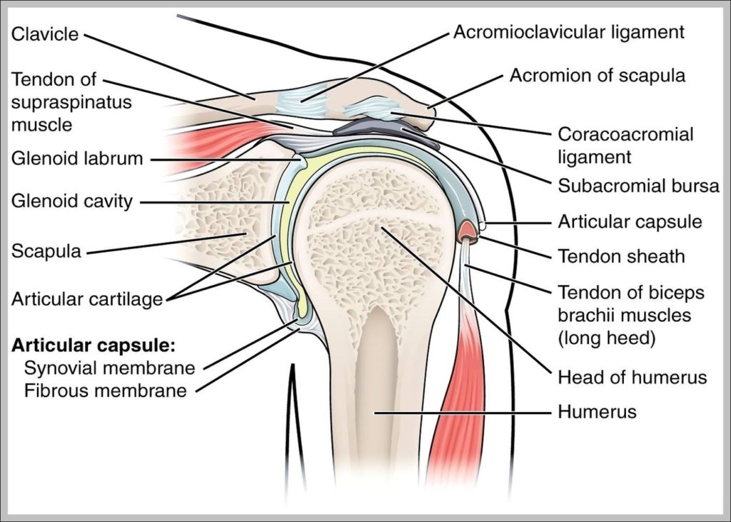
Shoulder Ligaments. Ligaments are soft tissue structures that connect bones to bones. There are several important ligaments in the shoulder. Glenohumeral Ligaments (GHL): A joint capsule is a watertight sac that surrounds a joint. In the shoulder, the joint capsule is formed by a group of ligaments that connect the humerus to the glenoid.
A joint capsule is a watertight sac that surrounds a joint. In the shoulder, the joint capsule is formed by a group of ligaments that connect the humerus to the glenoid. These ligaments are the main source of stability for the shoulder. They are the superior, middle and inferior glenohumeral ligaments.

Ligaments Of The Shoulder Joint Image Diagram - Ligaments Of The Shoulder Joint Image Chart - Human anatomy diagrams and charts explained. This anatomy system diagram depicts Ligaments Of The Shoulder Joint Image with parts and labels. Best diagram to help learn about health, human body and medicine.

Ligaments Of The Shoulder Joint Image

Ligaments In The Hand Image

In purely anatomical terms, only those ligaments that connect the bones of the hand to each other are properly called hand ligaments. The ligaments of the fingers and those that connect the hand to the wrist and forearm are not considered hand ligaments. The hand ligaments connect the bones of the hand.
The hand and wrist have many small ligaments that hold the bones together. Dorsal ligaments are on the back of the hand and include the carpometacarpal ligaments, dorsal metacarpal ligaments, dorsal intercarpal ligaments, dorsal radiocarpal ligaments, ulnar collateral ligament and the radial collateral ligament.

Ligaments In The Hand Image Diagram - Ligaments In The Hand Image Chart - Human anatomy diagrams and charts explained. This anatomy system diagram depicts Ligaments In The Hand Image with parts and labels. Best diagram to help learn about health, human body and medicine.

Ligaments In The Hand Image

Foot Ligaments Image

Because there are so many bones in the foot, there are also numerous ligaments connecting them. Some of the main ligaments in the foot are: Plantar fascia ligament: Runs underneath your foot from the heel to the base of your toes.
While the bones in the foot can fracture and the foot’s muscles can experience strains, one of the more common and potentially serious foot injuries are torn ligaments. Ligaments are the strong and flexible tissues that hold the bones throughout your body together; when a ligament tears, the resulting injury is often referred to as a sprain.

Foot Ligaments Image Diagram - Foot Ligaments Image Chart - Human anatomy diagrams and charts explained. This anatomy system diagram depicts Foot Ligaments Image with parts and labels. Best diagram to help learn about health, human body and medicine.

Foot Ligaments Image

Hand Ligaments Image

In purely anatomical terms, only those ligaments that connect the bones of the hand to each other are properly called hand ligaments. The ligaments of the fingers and those that connect the hand to the wrist and forearm are not considered hand ligaments. The hand ligaments connect the bones of the hand.
Two large ligaments bind the bones of the hand and wrist to the radius, a bone of the forearm. The first is on the back of the hand and is called the dorsal radiocarpal ligament, and the second, on the palm side of the hand, is called the palmar radiocarpal ligament.

Hand Ligaments Image Diagram - Hand Ligaments Image Chart - Human anatomy diagrams and charts explained. This anatomy system diagram depicts Hand Ligaments Image with parts and labels. Best diagram to help learn about health, human body and medicine.

Hand Ligaments Image

Ankle Ligaments

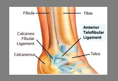
Ligaments of the ankle. Lateral ankle ligaments. The lateral side of the ankle has three supporting ligaments: the anterior talofibular ligament (ATFL), the posterior talofibular ligament (PTFL) and the calcaneofibular ligament(CFL). The three ligaments are together called the Lateral Collateral Ligament Complex.
Keywords: Ankle anatomy, Lateral collateral ligament, Medial collateral ligament, Ankle impingement, Ankle sprain Introduction Despite the fact that the ankle ligaments are prone to injury during the fast majority of sports, literature focusing on the ankle ligaments is rare.
On the outer side of your foot, you have several ligaments. These include the anterior talofibular ligament (ATFL) and the calcaneofibular ligament (CFL). These help keep your ankle and foot steady when you walk. If you have had repeated ankle sprains or if you have certain foot deformities, your ligaments can start to get weak and loose.

Ankle Ligaments Diagram - Ankle Ligaments Chart - Human anatomy diagrams and charts explained. This anatomy system diagram depicts Ankle Ligaments with parts and labels. Best diagram to help learn about health, human body and medicine.

ankle ligaments