Iliac Artery Anatomy Image

The common iliac arteries originate from the abdominal aorta. The internal iliac artery supplies the peritoneum, gluteal region and the walls and viscera of the pelvis. This article will discuss the anatomical course, anatomical relations and branches of the internal iliac artery as well its differences in the fetus.
One example is a common iliac artery aneurysm, which causes ballooning of the artery and can lead to rupture. The common iliac arteries are a link between the aorta and the arteries of the pelvis and lower limbs. There are two common iliac arteries that split off from the abdominal aorta (which moves blood from the heart).

Iliac Artery Anatomy Image Diagram - Iliac Artery Anatomy Image Chart - Human anatomy diagrams and charts explained. This anatomy system diagram depicts Iliac Artery Anatomy Image with parts and labels. Best diagram to help learn about health, human body and medicine.

Iliac Artery Anatomy Image

Common Iliac Vein Image

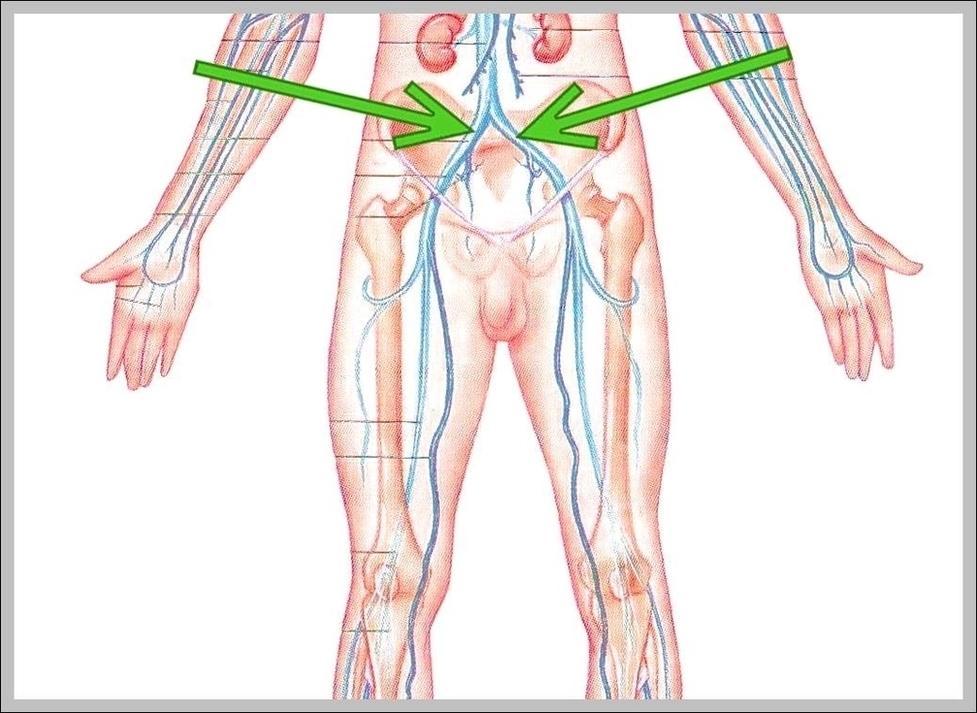
Common iliac vein. The common iliac vein is formed by the unification of the internal and external iliac veins. The external iliac vein drains the lower limb, and the internal iliac vein drains the gluteal region and pelvic viscera. The unification of the two common iliac veins forms the inferior vena cava.
Iliac Vein Compression Syndrome. Left common iliac vein stenosis frequently occurs where the vein crosses beneath the right common iliac artery. Chronic, repetitive compression at this site causes fibrosis of the vein, with synechiae and spurs that result in stenosis or even occlusion of the lumen.

Common Iliac Vein Image Diagram - Common Iliac Vein Image Chart - Human anatomy diagrams and charts explained. This anatomy system diagram depicts Common Iliac Vein Image with parts and labels. Best diagram to help learn about health, human body and medicine.

Common Iliac Vein Image

Iliac Node Image

Iliac lymph nodes. They are located above the pelvis and receive drainage from the sacral, external iliac and internal iliac groups of lymph nodes. They mainly drain lymph into the caval and aortic groups of lymph nodes. Some direct lymph drainage into the common iliac nodes occurs from some pelvic organs, for example,…
Iliac lymph nodes The location and number of several groups and sub-groups of lymph nodes receiving lymph drainage from the pelvic organs vary but four main groups are well known and can be described as follows: External iliac nodes: These group of lymph nodes are located above the pelvic brim, along the external iliac vessels.
They form a cluster around the anterior and posterior divisions of the internal iliac artery and the origins of the gluteal arteries. They receive drainage from the inferior pelvic viscera, deep perineum, and gluteal region and like the external iliac nodes, drain into the common iliac nodes.

Iliac Node Image Diagram - Iliac Node Image Chart - Human anatomy diagrams and charts explained. This anatomy system diagram depicts Iliac Node Image with parts and labels. Best diagram to help learn about health, human body and medicine.

Iliac Node Image