Cranial Nerve X Image

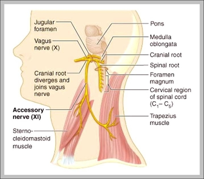
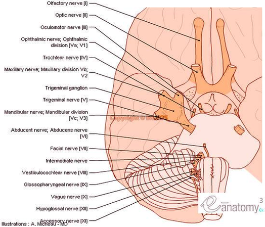
The cranial nerves are a set of twelve nerves that originate in the brain. Each has a different function for sense or movement. The functions of the cranial nerves are sensory, motor, or both:
Magnetic resonance imaging (MRI) is considered the gold standard in the study of the cranial nerves. Computed tomography (CT) allows, usually, an indirect view of the nerve and is useful to demonstrate the intraosseous segments of cranial nerves, the foramina through which they exit skull base and their pathologic changes.

Cranial Nerve X Image Diagram - Cranial Nerve X Image Chart - Human anatomy diagrams and charts explained. This anatomy system diagram depicts Cranial Nerve X Image with parts and labels. Best diagram to help learn about health, human body and medicine.

Cranial Nerve X Image

Cranial Nerves Anatomy Brainstem Human Body En Medical

Cranial Nerves Anatomy Brainstem Human Body En Medical Diagram - Cranial Nerves Anatomy Brainstem Human Body En Medical Chart - Human anatomy diagrams and charts explained. This anatomy system diagram depicts Cranial Nerves Anatomy Brainstem Human Body En Medical with parts and labels. Best diagram to help learn about health, human body and medicine.

cranial nerves anatomy brainstem human body en medical

Cranial Nerves Anatomy Brainstem Human Body En Large Photo

Cranial Nerves Anatomy Brainstem Human Body En Large Photo Diagram - Cranial Nerves Anatomy Brainstem Human Body En Large Photo Chart - Human anatomy diagrams and charts explained. This anatomy system diagram depicts Cranial Nerves Anatomy Brainstem Human Body En Large Photo with parts and labels. Best diagram to help learn about health, human body and medicine.

cranial nerves anatomy brainstem human body en large photo

Cranial Bones Diagram

Your cranial bones are eight bones that make up your cranium, or skull, which supports your face and protects your brain. We’ll go over each of these bones and where they’re located.
Cranial Floor View of the Skull Bones: (os frontale) Major Sutures of the Skull Sutures are junctions between adjacent bones of the skull which are rigidly held together by fibrous connective tissue. Fissures, foramina and markings of the base of the skull The cranial floor bones support the inferior part of skull.
Skull Features ©Sheri Amsel www.exploringnature.org frontal bone nasal bones temporal bone zygomatic arch (cheek bone) maxilla mandible external auditory meatus (ear canal) occipital bone frontal bone parietal bone mandible maxilla

Cranial Bones Diagram Diagram - Cranial Bones Diagram Chart - Human anatomy diagrams and charts explained. This anatomy system diagram depicts Cranial Bones Diagram with parts and labels. Best diagram to help learn about health, human body and medicine.

cranial bones diagram