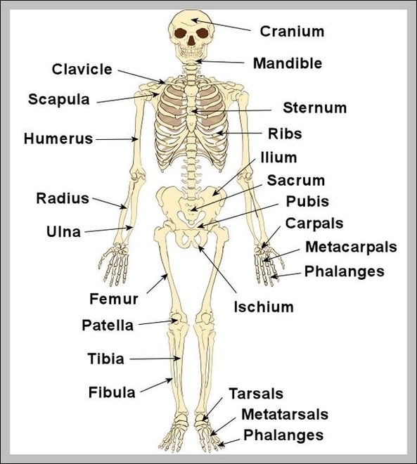
Human Bone Structure The human bone structure, also known as the skeletal system, serves as a framework for the body, providing support, protection, and enabling motion. It consists of many…
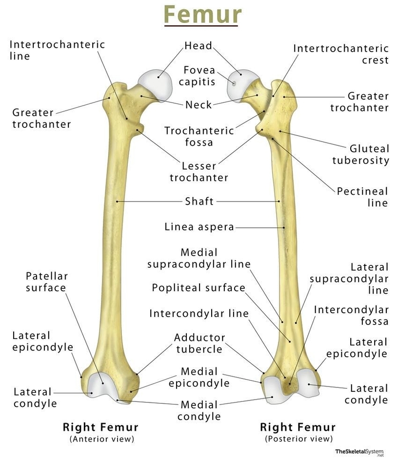
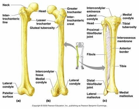
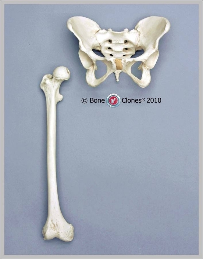
The femur, also known as the thigh bone, is the longest and strongest bone in the human body. It plays a crucial role in supporting body weight and facilitating movement.…
"Drag Each Label To The Appropriate Bone Marking" is a common exercise in anatomy and physiology courses, particularly in the study of the skeletal system. This activity involves identifying various…
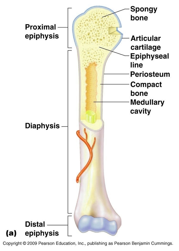
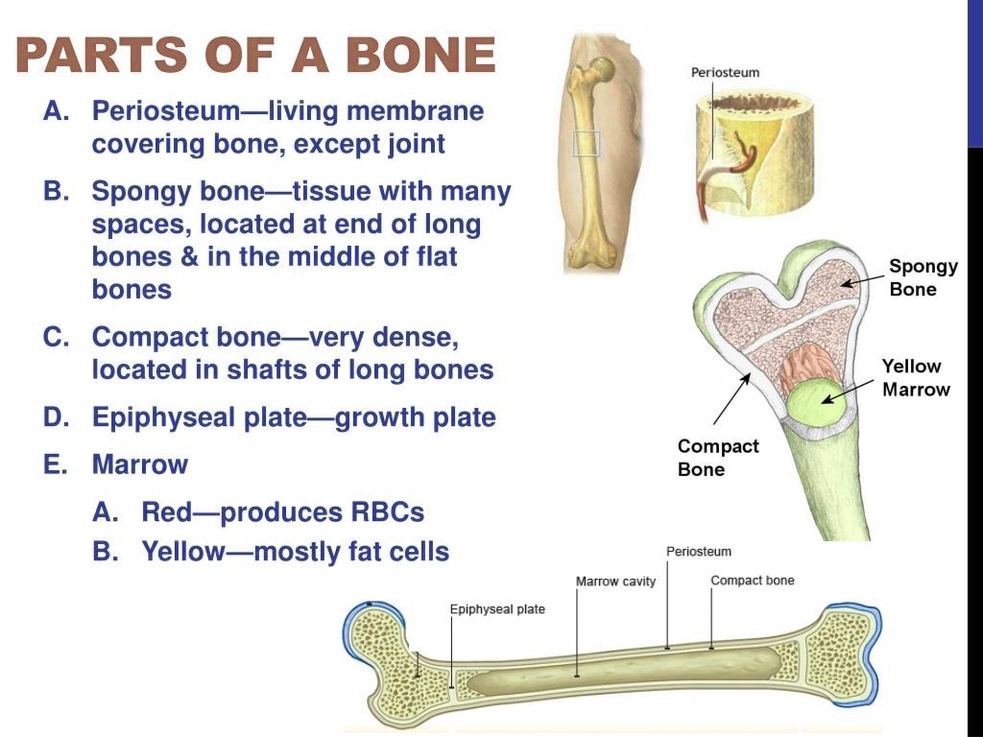
Internal Structure of Long Bones Long bones, as the name suggests, are longer than they are wide. They are one of the types of bones classified based on their shape,…
The human leg, a marvel of biological engineering, is a complex structure composed of numerous bones that work in harmony to provide support and mobility. Femur (Thighbone) The femur, or…
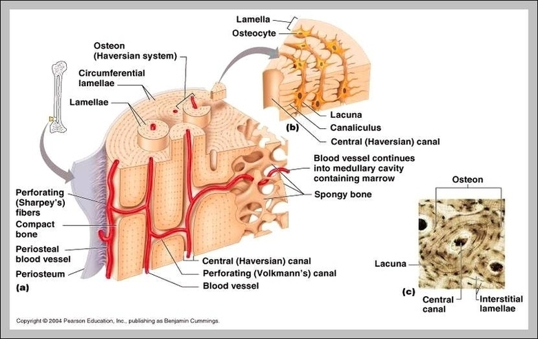
Bone Parts Bones are the structural framework of the body, providing support, protection, and facilitating movement. They are composed of several parts, each with a unique function: 1. Osteon: The…
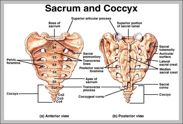
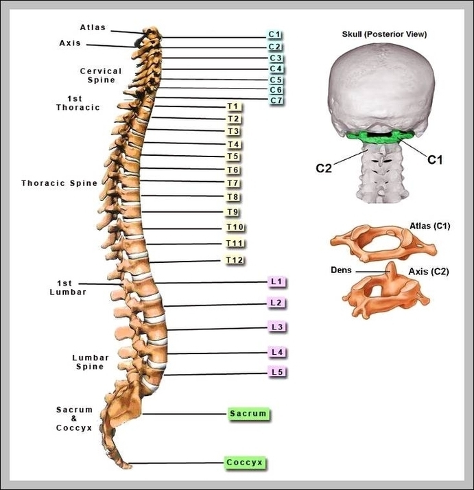
The coccyx is a triangular arrangement of bone that makes up the very bottom portion of the spine below the sacrum. It represents a vestigial tail, hence the common term…
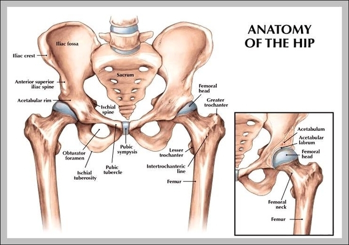
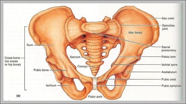
4,902 hip anatomy stock photos and images available, or search for human hip anatomy or hip anatomy illustration to find more great stock photos and pictures. General Hip Anatomy. The…
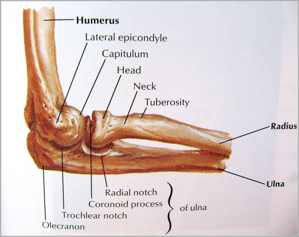
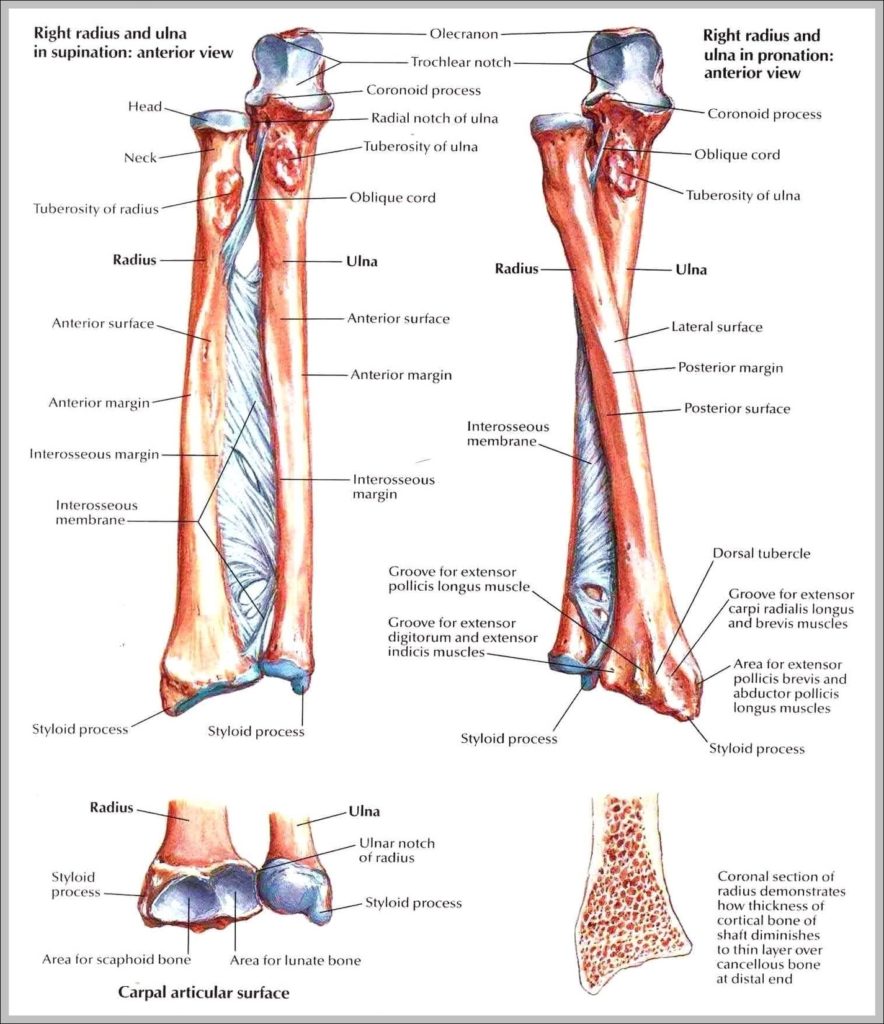
The Anatomy of the Elbow The elbow is a hinged joint made up of three bones, the humerus, ulna, and radius. The ends of the bones are covered with cartilage.…
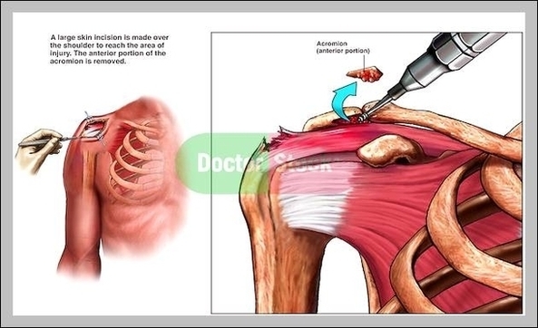
The acromion is a continuation of the scapular spine, and hooks over anteriorly. It articulates with the clavicle (collar bone) to form the acromioclavicular joint. [edit on Wikidata] In human…
1,047 hip bone stock photos and images available, or search for hip bone icon or hip bone 3d to find more great stock photos and pictures. Bony Structures of the…
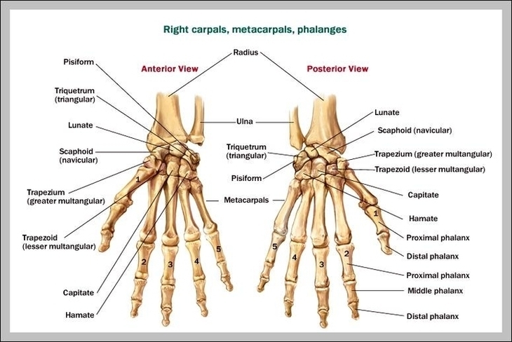
2,316 wrist bone stock photos and images available, or search for wrist xray or wrist pain to find more great stock photos and pictures. Film X-ray wrist radiograph show carpal…
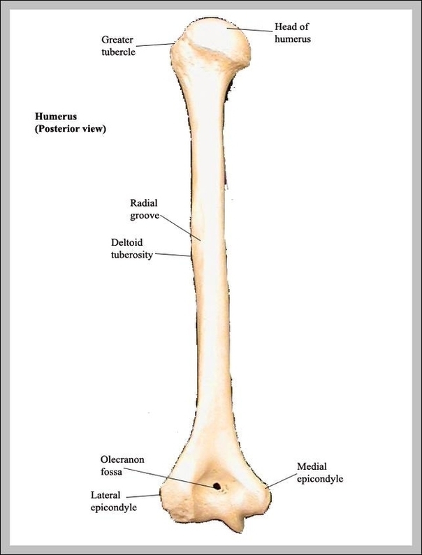
3,852 arm bone stock photos and images available or search for broken arm bone or human arm bone to find more great stock photos and pictures. Hemophilic Arthropathy Of The…
Microscopic Structure of Bones. The skeleton makes up about 30-40% of an adult’s body mass. The skeleton’s mass is made up of nonliving bone matrix and many tiny bone cells.…
67,060 human bone stock photos and images available, or search for human bone marrow or human bone structure to find more great stock photos and pictures. Abstract image human body…