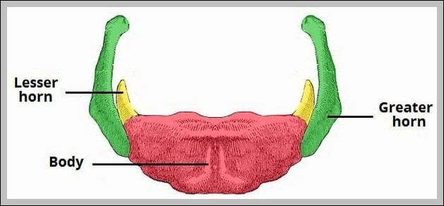
Human Body Anatomy Diagrams Described In Detail

Human Body Anatomy

The human body is a complex and intricate system, composed of many different types of cells that together create tissues, organs, and organ systems. These components ensure homeostasis and the viability of the human body.

1. Basic Structure

The human body consists of the head, neck, torso (which includes the thorax and abdomen), arms and hands, legs and feet. The skeleton, composed of cartilage and bone, gives the body its shape.

2. Cells and Tissues

The body contains trillions of cells, the fundamental unit of life. These cells sit in an extracellular matrix that consists of proteins such as collagen, surrounded by extracellular fluids. Not all parts of the body are made from cells. Some parts are non-cellular material such as bone and connective tissue.

3. Organ Systems

The human body is organized into several major organ systems. Each system has a specific function and is made up of specific organs and tissues.

– Cardiovascular System: This system includes the heart and blood vessels. It circulates blood throughout the body, delivering oxygen and nutrients to the cells.
– Digestive System: It breaks down food into nutrients that can be absorbed and used by the body.
– Endocrine System: It consists of glands that produce hormones, which regulate many body functions like growth and metabolism.
– Renal System: It filters the blood and removes waste products through urine.
– Muscular System: It allows movement and provides support to the body.
– Nervous System: It controls and coordinates body activities and senses the environment.
– Reproductive System: It allows humans to reproduce.
– Respiratory System: It brings in oxygen and expels carbon dioxide.
– Skeletal System: It provides structure, protects organs, and enables movement.

4. Biochemical Constituents

The human body is composed of elements including hydrogen, oxygen, carbon, calcium, and phosphorus. These elements reside in trillions of cells and non-cellular components of the body. The main electrolytes in body water outside cells are sodium and chloride, whereas within cells it is potassium and other phosphates.

5. Development and Aging

The human body undergoes various stages of development, from conception through old age. This includes prenatal development, growth, and aging.

In conclusion, the human body is a marvel of biological engineering, with each part working in harmony to ensure the survival and well-being of the individual. Understanding its anatomy and physiology is crucial for medical and health-related fields.

Human Body Anatomy Diagrams Described In Detail Diagram - Human Body Anatomy Diagrams Described In Detail Chart - Human anatomy diagrams and charts explained. This anatomy system diagram depicts Human Body Anatomy Diagrams Described In Detail with parts and labels. Best diagram to help learn about health, human body and medicine.

Human Body Anatomy Diagrams Described In Detail

Ventral Body Cavity Anatomy

Ventral Body Cavity Anatomy

The ventral body cavity, also known as the ventral cavity, is a human body cavity located at the anterior (front) aspect of the human body. It is a fluid-filled space that surrounds the organs on the ventral side of humans and other tetrapods. This cavity is one of two main cavities, the other being the dorsal cavity.

The ventral body cavity is divided into two main parts: the thoracic cavity and the abdominopelvic cavity. These cavities are separated by the diaphragm, a thin muscle that helps control the expansion and contraction of the lungs.

Thoracic Cavity

The thoracic cavity contains the heart, lungs, breast tissue, thymus gland, and blood vessels. It is further divided into separate parts. Two cavities, the left and right pleural cavities, hold the lungs. A central membrane, the mediastinum, divides these two chambers. The heart sits within the pericardial cavity.

Abdominopelvic Cavity

The abdominopelvic cavity is further divided into the abdominal cavity and pelvic cavity. The abdominal cavity contains digestive organs, spleen, and the kidneys. The pelvic cavity contains the urinary bladder, internal reproductive organs, and rectum.

There are two methods for dividing the abdominopelvic cavity. The clinical method, used by physicians and nurses, utilizes four sections called quadrants: the right upper quadrant, the left upper quadrant, the right lower quadrant, and the left lower quadrant. The second method for dividing the abdominopelvic cavity is preferred by anatomists. This method divides the cavity into nine regions.

Function of the Ventral Cavity

The ventral cavity has several important functions relating to the organs housed within it. First and foremost, the cavity protects the organs inside from shock damage as the organism moves through the world. The space and fluid around the organs ensure that any impacts incurred by the organism will not be transferred onto the organs.

A function which is used more by animals with lungs is expansion, or the ability of the ventral cavity to change shape, allowing the expansion of certain organs. In humans, the ventral cavity must expand in several places to allow for various organs to expand and change shape.

Both the organs and the ventral cavity walls are lined with serosa, a thin membrane which separates the cavity from the inside of the body. The organs are also lined with a peritoneum, protecting them from rubbing on the inside of the cavity. Together, this creates a system which allows the organs to slide seamlessly past one another as the body moves about.

In conclusion, the ventral body cavity plays a crucial role in protecting and accommodating the organs within the human body. Its structure and function are integral to the overall functioning of the human body..

Ventral Body Cavity Anatomy Diagram - Ventral Body Cavity Anatomy Chart - Human anatomy diagrams and charts explained. This anatomy system diagram depicts Ventral Body Cavity Anatomy with parts and labels. Best diagram to help learn about health, human body and medicine.

Ventral Body Cavity Anatomy

Major Skeletal Muscles Of The Body

Major Skeletal Muscles Of The Body
keletal muscles, which make up between 30 to 40% of your total body mass, are the muscles that connect to your bones and allow you to perform a wide range of movements and functions. They are voluntary muscles, meaning you control how and when they work. There are more than 600 muscles in the human body.

Function and Structure
keletal muscles serve a variety of functions, including chewing and swallowing, expanding and contracting your chest cavity for respiration, maintaining body posture, moving the bones in different parts of your body, and protecting joints and holding them in place.

Each muscle can contain thousands of fibers. Different types of sheaths, or coverings, surround the fibers: Epimysium is the outermost layer of tissue surrounding the entire muscle.

Interactions of Skeletal Muscles

To move the skeleton, the tension created by the contraction of the fibers in most skeletal muscles is transferred to the tendons. The tendons are strong bands of dense, regular connective tissue that connect muscles to bones. The bone connection is why this muscle tissue is called skeletal muscle.

The moveable end of the muscle that attaches to the bone being pulled is called the muscle’s insertion, and the end of the muscle attached to a fixed (stabilized) bone is called the origin.

Prime Movers and Synergists

Although a number of muscles may be involved in an action, the principal muscle involved is called the prime mover, or agonist. A muscle with the opposite action of the prime mover is called an antagonist. Antagonists play two important roles in muscle function: They maintain body or limb position, such as holding the arm out or standing erect. They control rapid movement, as in shadow boxing without landing a punch or the ability to check the motion of a limb.

Major Muscles of Upper Limb

– Deltoid: shoulder abduction, flexion and extension
– Biceps brachii: flexes elbow
– Triceps brachii: extends forearm

Major Muscles of Lower Limb

– Gluteus maximus: external rotation and extension of the hip joint
– Sartorius: lateral rotation and abduction of thigh; flexion and medial rotation of leg

Facial Muscles

There are also skeletal muscles that do not pull against the skeleton for movements. For example, there are the muscles that produce facial expressions. The insertions and origins of facial muscles are in the skin, so that certain individual muscles contract to form a smile or frown, form sounds or words, and raise the eyebrows.

In conclusion, skeletal muscles are a vital part of your musculoskeletal system. They are responsible for the movement of the human body, allowing us to perform a wide range of actions and functions. Understanding the structure and function of these muscles is crucial for understanding human anatomy and physiology.

Major Skeletal Muscles Of The Body Diagram - Major Skeletal Muscles Of The Body Chart - Human anatomy diagrams and charts explained. This anatomy system diagram depicts Major Skeletal Muscles Of The Body with parts and labels. Best diagram to help learn about health, human body and medicine.

Major Skeletal Muscles Of The Body

Types Of Muscle Tissue In Human Body Explained

Types of Muscle Tissue in the Human Body

Muscle tissue is a specialized tissue found in animals which functions by contracting, thereby applying forces to different parts of the body. There are three types of muscle tissue: skeletal, cardiac, and smooth.

1. Skeletal Muscle: This type of tissue is found in skeletal muscles and is responsible for the voluntary movements of bones. It is a type of striated muscle, meaning clear bands can be seen in it under a microscope. These tiny light and dark bands are highly organized bundles of actin, myosin, and associated proteins. These organized bundles allow striated muscle to contract quickly and release quickly. Skeletal muscle tissue can be controlled voluntarily, by the brain.

2. Cardiac Muscle: This is the specialized muscle of the heart. It is also a type of striated muscle, but unlike skeletal muscle, it is formed of short, uninucleate, branching myocytes which are connected at intercalated discs. These junctures help cardiac muscle to contract as one and provide a rapid and coordinated contraction to move blood.

3. Smooth Muscle: This is a non-striated muscle, formed of short, uninucleate, spindle-shaped myocytes. It is located in the walls of internal organs, blood vessels, etc. Unlike cardiac and skeletal muscle tissue, smooth muscle tissue has no striations. The fibers of myosin and actin in smooth muscle fiber are not nearly as organized as in the other types of muscle tissue.

Each type of muscle tissue in the human body has a unique structure and a specific role. Skeletal muscle moves bones and other structures. Cardiac muscle contracts the heart to pump blood. Smooth muscle can be found in the walls of hollow organs, such as the intestines, uterus, and stomach.

Muscle tissue has four main properties: Excitability (an ability to respond to stimuli), Contractibility (an ability to contract), Extensibility (an ability to be stretched without tearing), and Elasticity (an ability to return to its normal shape). Through these properties, the muscular system as a whole performs several important functions. These include the production of force and movement, support of body stature and position, stability of joints, production of body heat (to maintain normal body temperature), as well as, provision of form to the body.

In conclusion, muscle tissues play a crucial role in the functioning of the human body. Their ability to contract and relax enables us to move, pump blood, and perform various other vital functions. Understanding the different types of muscle tissues and their functions can provide valuable insights into how our bodies work..

Types Of Muscle Tissue In Human Body Explained Diagram - Types Of Muscle Tissue In Human Body Explained Chart - Human anatomy diagrams and charts explained. This anatomy system diagram depicts Types Of Muscle Tissue In Human Body Explained with parts and labels. Best diagram to help learn about health, human body and medicine.

Types Of Muscle Tissue In Human Body Explained

Human Body Systems Represented

The human body is a complex biological machine, composed of various systems that work together to sustain life. Here’s an overview of the main body systems:

1. Musculoskeletal System: Provides mechanical support, posture, and locomotion. It consists of bones, cartilages, and muscles.

2. Cardiovascular System: Transports oxygen, nutrients, and hormones throughout the body and eliminates cellular metabolic waste. It includes the heart, arteries, and veins.

3. Respiratory System: Facilitates the exchange of oxygen and carbon dioxide between the body and air, regulates acid-base balance, and enables phonation. It includes organs like the lungs and trachea.

4. Nervous System: Initiates and regulates vital body functions, sensation, and body movements. It is divided into the central nervous system (brain and spinal cord) and the peripheral nervous system (nerves outside the brain and spinal cord).

5. Digestive System: Mechanically and chemically degrades food for absorption into the body and use as energy. It includes the mouth, esophagus, stomach, and intestines.

6. Urinary System: Filters blood and eliminates unnecessary compounds and waste by producing and excreting urine. It includes the kidneys, ureters, bladder, and urethra.

7. Endocrine System: Produces hormones to regulate a wide variety of bodily functions, such as the menstrual cycle and sugar levels. It includes glands like the pituitary, thyroid, and adrenal glands.

8. Lymphatic System: Drains excess tissue fluid and defends the body against pathogens. It includes the lymph nodes, lymphatic vessels, and lymph.

9. Reproductive System: Produces reproductive cells and contributes to the reproduction process. It differs between males and females, with organs like the ovaries and uterus in females, and the testes and prostate in males.

10. Integumentary System: Provides physical protection of the body surface, sensory reception, and vitamin synthesis. It includes the skin and its appendages like hair, nails, sweat glands, and oil glands.

Each system has its unique functions, organs, and histology. Some organs may be part of more than one body system if they serve more than one function. These systems work in harmony to maintain the body’s overall health and functionality. Understanding these systems and how they interact is fundamental to understanding human biology..

Human Body Systems Represented Diagram - Human Body Systems Represented Chart - Human anatomy diagrams and charts explained. This anatomy system diagram depicts Human Body Systems Represented with parts and labels. Best diagram to help learn about health, human body and medicine.

Human Body Systems Represented

Labeled Anatomy Human Body

The human body is a complex and intricate system, composed of various organs, tissues, and cells, all working in harmony to maintain life. Here’s an overview of the labeled anatomy of the human body:

1. Vital Organs
These are organs that a person needs to survive. Any problem with these organs can quickly become life-threatening:

– Brain: The body’s control center, creating, sending, and processing nerve impulses, thoughts, emotions, physical sensations, and more.
– Heart: The most important organ of the circulatory system, which helps deliver blood to the body.
– Lungs: Essential for respiration, allowing oxygen in the air to be taken into the body while also enabling the body to get rid of carbon dioxide.
– Liver: Performs various functions such as detoxification, protein synthesis, and production of biochemicals necessary for digestion.
– Kidneys: Responsible for filtering waste products, excess water, and other impurities from the blood.

2. Non-Vital Organs
These organs are not necessary for survival, but they do have a role in bodily functions. Examples include the gallbladder, pancreas, and stomach.

3. Organ Systems
These are groups of organs that work together to perform complex functions. For example, the nervous system supports the brain and other organs.

4. Tissues, Cells, and Extracellular Materials
The human body is composed of living cells and extracellular materials, organized into tissues, organs, and systems.

5. Biochemical Constituents
The body’s biochemical constituents include proteins, carbohydrates, lipids, nucleic acids, vitamins, and hormones.

6. Development
The human body undergoes various stages of development, from conception through old age.

7. Sensory Reception
The human body has a complex sensory reception system that allows us to interact with our environment.

8. Muscular and Skeletal Systems
The human muscle system allows for movement and the skeletal system provides the structural framework for the body.

9. Reproductive System
The human reproductive system enables reproduction and the continuation of our species.

10. Respiratory System
The human respiratory system allows for the intake of oxygen and the expulsion of carbon dioxide.

11. Endocrine System
The human endocrine system regulates the body’s growth, metabolism, and sexual development and function.

12. Digestive System
The human digestive system breaks down food into nutrients that the body can use.

13. Cardiovascular System
The cardiovascular system circulates blood throughout the body, delivering oxygen and nutrients to the cells.

In conclusion, the human body is a marvel of biological engineering, with each part playing a crucial role in maintaining overall health and function. Understanding the labeled anatomy of the human body is fundamental to appreciating the complexity and beauty of human life..

Labeled Anatomy Human Body Diagram - Labeled Anatomy Human Body Chart - Human anatomy diagrams and charts explained. This anatomy system diagram depicts Labeled Anatomy Human Body with parts and labels. Best diagram to help learn about health, human body and medicine.

Labeled Anatomy Human Body

Human Body Tissues Explained

Human Body Tissues Explained

The human body is a complex structure composed of four primary tissue types: epithelial, connective, muscle, and nervous. These tissues, each with their unique structure and function, work together to maintain the overall health and functionality of the body.

1. Epithelial Tissue: This tissue type covers the exterior surfaces of the body, lines internal cavities and passageways, and forms certain glands. It is made of layers of cells that cover the surfaces of the body that come into contact with the exterior world.

2. Connective Tissue: As its name implies, connective tissue binds the cells and organs of the body together. It provides support and structure to the body and is essential for our overall health and well-being.

3. Muscle Tissue: Muscle tissue contracts forcefully when excited, providing movement. It is crucial for our ability to move and perform physical tasks.

4. Nervous Tissue: Nervous tissue is excitable, allowing for the generation and propagation of electrochemical signals in the form of nerve impulses that communicate between different regions of the body. It is essential for our ability to sense and respond to our environment.

These primary tissue types originate from the three germ layers formed during embryonic development: ectoderm (outer), mesoderm (middle), and endoderm (inner). The cells composing a tissue share a common embryonic origin.

The organization of these tissues into organs and organ systems allows the body to function as a whole. For example, the digestive system is responsible for taking in and processing food, while the respiratory system—working with the circulatory system—is responsible for taking up oxygen and getting rid of carbon dioxide. The muscular and skeletal systems are crucial for movement; the reproductive system handles reproduction; and the excretory system gets rid of metabolic waste.

In conclusion, understanding the various primary tissue types present in the human body is essential for understanding the structure and function of organs which are composed of two or more primary tissue types. The survival of the organism depends on the integrated activity of all the organ systems, often coordinated by the endocrine and nervous systems..

Human Body Tissues Explained Diagram - Human Body Tissues Explained Chart - Human anatomy diagrams and charts explained. This anatomy system diagram depicts Human Body Tissues Explained with parts and labels. Best diagram to help learn about health, human body and medicine.

Human Body Tissues Explained

Human Body Photo Image

Human body photo image showing various parts of human body anatomy.

Human Body Photo Image Diagram - Human Body Photo Image Chart - Human anatomy diagrams and charts explained. This anatomy system diagram depicts Human Body Photo Image with parts and labels. Best diagram to help learn about health, human body and medicine.

Human Body Photo Image

Internal Body Anatomy Image

Internal Body Anatomy Image Diagram - Internal Body Anatomy Image Chart - Human anatomy diagrams and charts explained. This anatomy system diagram depicts Internal Body Anatomy Image with parts and labels. Best diagram to help learn about health, human body and medicine.

Internal Body Anatomy Image

Appendix Location On Body Image

Labeled Diagram of the Human Lungs Lungs are an excellent example of how several tissues can be compactly arranged, yet providing a large surface area for gaseous exchange. The current article provides a labeled diagram of the human lungs as well as a description of the parts and their functions.
7,786 human lung anatomy stock photos and images available, or start a new search to explore more stock photos and images.

Appendix Location On Body Image Diagram - Appendix Location On Body Image Chart - Human anatomy diagrams and charts explained. This anatomy system diagram depicts Appendix Location On Body Image with parts and labels. Best diagram to help learn about health, human body and medicine.

Appendix Location On Body Image

Diagram Of The Body Systems Image

A human body diagram is a representation that outlines the major structures within the human body. This diagram should include a drawing or outline of the skeletal system and internal organs within the body.
Luckily, though, you can now download a free body diagram template and use it as a visual aid in the classroom. The students of today need understanding, so, you cannot teach a topic like this without a body diagram template.

Diagram Of The Body Systems Image Diagram - Diagram Of The Body Systems Image Chart - Human anatomy diagrams and charts explained. This anatomy system diagram depicts Diagram Of The Body Systems Image with parts and labels. Best diagram to help learn about health, human body and medicine.

Diagram Of The Body Systems Image

Diagram Of Human Organs In The Body Female Image

507 female body diagram stock photos and images available, or search for body silhouette or human body diagram to find more great stock photos and pictures. The Principal Glands Of The Female And Male Human Endocrine Systems.
7,751 organs of the human body diagram stock illustrations and vector graphics available royalty-free, or start a new search to explore more great stock images and vector art. internal organs and circulatory system Vector isolated illustration of human internal organs and circulatory system in man body.

Diagram Of Human Organs In The Body Female Image Diagram - Diagram Of Human Organs In The Body Female Image Chart - Human anatomy diagrams and charts explained. This anatomy system diagram depicts Diagram Of Human Organs In The Body Female Image with parts and labels. Best diagram to help learn about health, human body and medicine.

Diagram Of Human Organs In The Body Female Image

Body Anatomy Liver Image

4,742 human liver anatomy stock photos and images available, or start a new search to explore more stock photos and images.
The liver is a large, meaty organ that sits on the right side of the belly. Weighing about 3 pounds, the liver is reddish-brown in color and feels rubbery to the touch. Normally you can’t feel the liver, because it’s protected by the rib cage. The liver has two large sections, called the right and the left lobes.

Body Anatomy Liver Image Diagram - Body Anatomy Liver Image Chart - Human anatomy diagrams and charts explained. This anatomy system diagram depicts Body Anatomy Liver Image with parts and labels. Best diagram to help learn about health, human body and medicine.

Body Anatomy Liver Image

Human Male Body Image

5,814 male human anatomy diagram stock photos and images available, or start a new search to explore more stock photos and images. Male silhouette (contour) on white background, vector. Male, female and children’s silhouette on white background,…
Skinny, muscular and fat physique. Healthy young man from front and back view in outline style. Male muscular body shapes linear illustration with the inscription: front and back. Male and female human vector silhouettes.

Human Male Body Image Diagram - Human Male Body Image Chart - Human anatomy diagrams and charts explained. This anatomy system diagram depicts Human Male Body Image with parts and labels. Best diagram to help learn about health, human body and medicine.

Human Male Body Image

Human Body Stomach Image

2,755 human stomach anatomy stock photos and images available or start a new search to explore more stock photos and images.
Next. The stomach is a muscular organ located on the left side of the upper abdomen. The stomach receives food from the esophagus. As food reaches the end of the esophagus, it enters the stomach …

Human Body Stomach Image Diagram - Human Body Stomach Image Chart - Human anatomy diagrams and charts explained. This anatomy system diagram depicts Human Body Stomach Image with parts and labels. Best diagram to help learn about health, human body and medicine.

Human Body Stomach Image

Image Of Human Body Image

Abstract image human body in the form of a starry sky or space, consisting of points, lines, and shapes in the form of planets, stars and the universe. Low poly vector background. human body anatomy stock illustrations Abstract image human body in the form of a starry sky or space,…
130,635 inside the human body stock photos and images available, or search for human anatomy or blood cells to find more great stock photos and pictures.

Image Of Human Body Image Diagram - Image Of Human Body Image Chart - Human anatomy diagrams and charts explained. This anatomy system diagram depicts Image Of Human Body Image with parts and labels. Best diagram to help learn about health, human body and medicine.

Image Of Human Body Image

Hinge Joints In The Body Image

A hinge joint is a common class of synovial joint that includes the ankle, elbow, and knee joints. Hinge joints are formed between two or more bones where the bones can only move along one axis to flex or extend.
The elbows, the knees, and the middle and end joints of the fingers are hinge joints. Hinge joints are prone to injury when lateral forces are applied to the joint. Many knee injuries occur in this manner, according to the National Institute of Arthritis and Musculoskeletal and Skin Diseases.

Hinge Joints In The Body Image Diagram - Hinge Joints In The Body Image Chart - Human anatomy diagrams and charts explained. This anatomy system diagram depicts Hinge Joints In The Body Image with parts and labels. Best diagram to help learn about health, human body and medicine.

Hinge Joints In The Body Image

Labeled Body Parts In Spanish Image

The names of body parts are used much the same as they are in Spanish as in English, but with one significant difference. In Spanish, names of parts of the body are frequently preceded by the definite article (el, la, los or las, meaning “the”) instead of possessive adjectives (such as mi for “my” and tu for “your”).
I broke my [body part]. (Ex. I broke my arm.) Me rompí [body part]. (Ex. Me rompí el brazo.) I cut my finger. Me corto el dedo. I have a pain/ache of [body part]. (Ex. I have a headache.)** Tengo dolor de [body part]. (Ex.Tengo dolor de cabeza.) I have black/blond/brown/red/gray hair.

Labeled Body Parts In Spanish Image Diagram - Labeled Body Parts In Spanish Image Chart - Human anatomy diagrams and charts explained. This anatomy system diagram depicts Labeled Body Parts In Spanish Image with parts and labels. Best diagram to help learn about health, human body and medicine.

Labeled Body Parts In Spanish Image

Pictures Of Bones In The Human Body Image

67,060 human bone stock photos and images available, or search for human bone marrow or human bone structure to find more great stock photos and pictures.
12,251 human body organs anatomy stock photos and images available, or start a new search to explore more stock photos and images.

Pictures Of Bones In The Human Body Image Diagram - Pictures Of Bones In The Human Body Image Chart - Human anatomy diagrams and charts explained. This anatomy system diagram depicts Pictures Of Bones In The Human Body Image with parts and labels. Best diagram to help learn about health, human body and medicine.

Pictures Of Bones In The Human Body Image

A Human Body Image

Abstract image of a human body in the form of a starry sky or space, consisting of points, lines, and shapes in the form of planets, stars and the universe. Low poly vector background. the human body stock illustrations
394,312 the human body stock photos and images available or search for the human body anatomy body or the human body photos to find more great stock photos and pictures.

A Human Body Image Diagram - A Human Body Image Chart - Human anatomy diagrams and charts explained. This anatomy system diagram depicts A Human Body Image with parts and labels. Best diagram to help learn about health, human body and medicine.

A Human Body Image