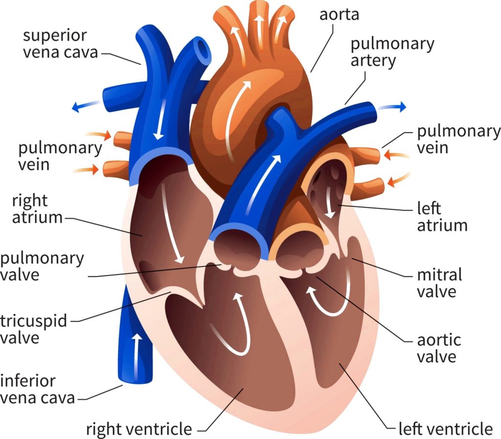
Posted inDiagrams
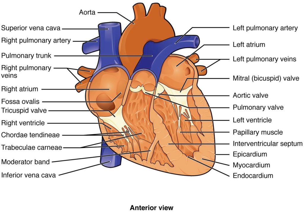
External Structure Of Heart Anatomy Diagram
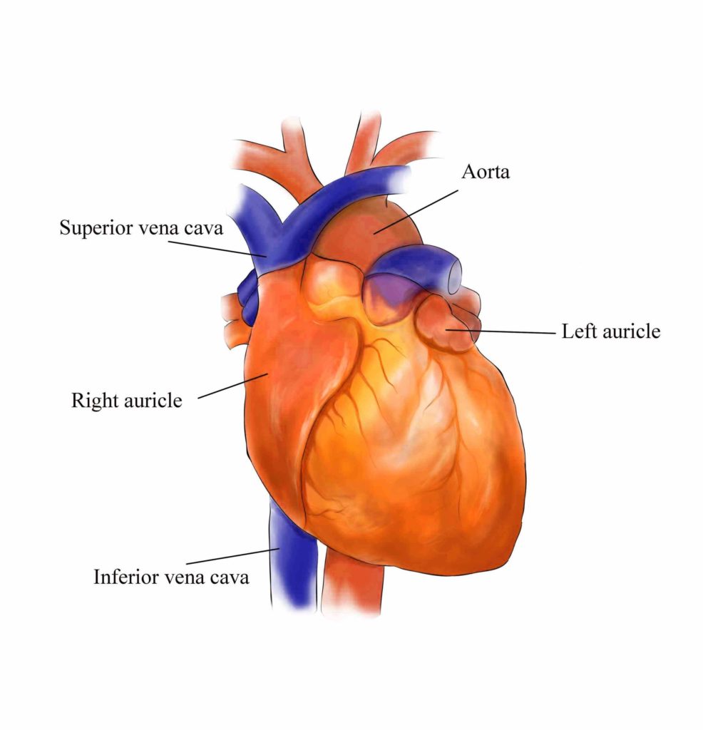
External Structure of the Heart Anatomy The heart, a muscular organ, is responsible for circulating blood throughout the body via the circulatory/vascular system. It is located in the middle mediastinum,…