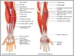
The antecubital fossa is bounded on the top by the skin and several veins and nerves, while it is bounded on the bottom by more muscles. The antecubital fossa houses several important structures. The radial nerve passes on the same side of the arm as the radius. The radial nerve supplies many of the muscles of the arm.
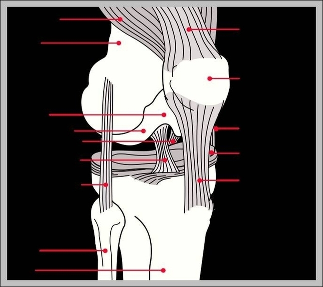
It arises from the distal part of the bone, below your biceps brachii muscle. It then courses down the front of your arm, over your elbow joint, and inserts on the coronoid process and tuberosity of your ulna. The brachialis muscle, along with the supinator muscle, makes up the floor of the cubital fossa of your elbow.
The brachialis is a muscle in the front of your elbow that flexes, or bends, the joint. It does this when your forearm is in a palm down, pronated, position.
Brachial Fossa Image
Posted inDiagrams
Brachial Fossa Image
Post navigation
Previous Post
 Pictures Of Female Anatomy Image
Next Post
Anatomy Lessons Image