This makes the ankle one of the most stable joints in the lower extremities. Here is a brief definition of each of the ankle bones: The tibia forms the inside (medial) portion of the ankle. The fibula forms the outside (lateral) portion of the ankle. The talus is also known as the ankle bone and is found underneath the tibia and fibula.
8,460 ankle anatomy stock photos and images available, or search for foot and ankle anatomy to find more great stock photos and pictures. The ankle joint, tendons of the ankle joint foot anatomy vector… bones of the foot and ankle joint medical vector illustration… Houman body parts flat line icons set.
8,460 ankle anatomy stock photos and images available, or search for foot and ankle anatomy to find more great stock photos and pictures. The ankle joint, tendons of the ankle joint foot anatomy vector… bones of the foot and ankle joint medical vector illustration… Houman body parts flat line icons set.
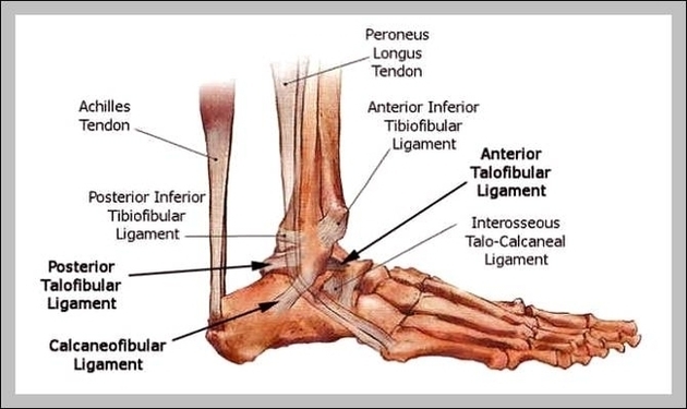
Ankle Bones Diagram Image
Posted inDiagrams
Ankle Bones Diagram Image
Post navigation
Previous Post
 Human Organ Picture Image
Next Post
Inside Body Picture Image