The angles of the ribs form the most posterior extent of the thoracic cage. In the anatomical position, the angles align with the medial border of the scapula. A shallow costal groove for the passage of blood vessels and a nerve is found along the inferior margin of each rib. The bony ribs do not extend anteriorly completely around to the sternum.
The thoracic cage consists of ribs and the sternum. It is a bony structure that protects the organs in the thoracic cavity. It includes 25 bones, i.e., 1 sternum and 24 ribs. A. Ribs: They are a cage-like formation of thin, flat and curved bones.
The potential for movement is related to the flexibility provided by the ribs and their joints. The thoracic cage is composed of the thoracic skeleton, which includes the sternum, 12 pairs of ribs and 12 thoracic vertebrae, associated with the costal cartilages and intervertebral discs, respectively.
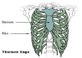
thoracic cage diagram
Posted inDiagrams
Thoracic cage diagram
Post navigation
Previous Post
Next Post
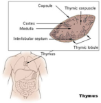
Thymus diagram