The wrist tendons are: Flexor carpi radialis: This tendon is one of two tendons that bend the wrist. It attaches to the base of the second and third hand bones.
Wrist MRI Anatomy: T1-weighted coronal view. Image 15. 9, Trapezium. 10, Pisiform. 14, Thenar mm. This webpage presents the anatomical structures found on wrist MRI. The wrist consists of multiple joints where the bones of the arm and hand meet to facilitate movement (1).
Wrist MRI Anatomy: T1-weighted coronal view. Image 15. 9, Trapezium. 10, Pisiform. 14, Thenar mm. This webpage presents the anatomical structures found on wrist MRI. The wrist consists of multiple joints where the bones of the arm and hand meet to facilitate movement (1).
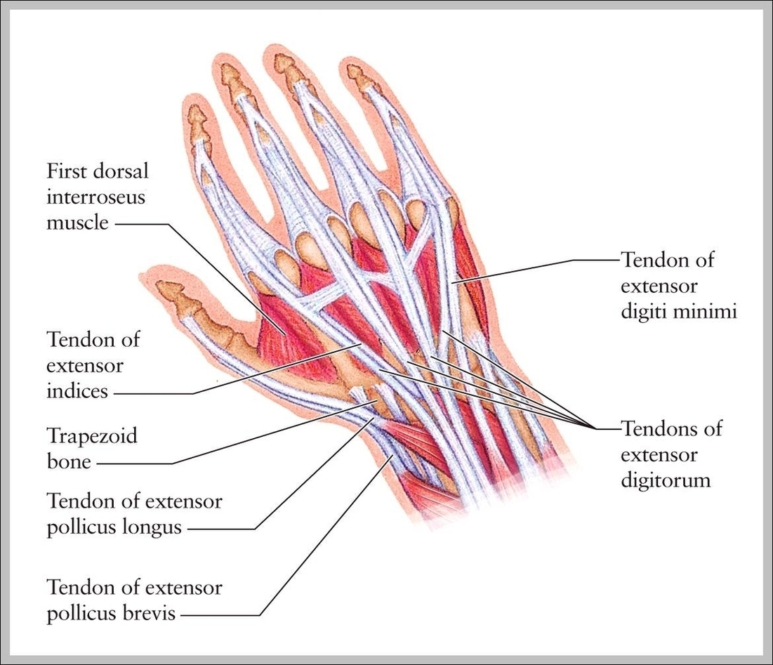
Wrist Tendons Anatomy Image
Posted inDiagrams
Wrist Tendons Anatomy Image
Post navigation
Previous Post
 How To Become A Home Health Aid Image
Next Post
Diagram Of Urinary System Image