
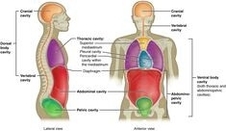
Ventral Body Cavity Anatomy
The ventral body cavity, also known as the ventral cavity, is a human body cavity located at the anterior (front) aspect of the human body. It is a fluid-filled space that surrounds the organs on the ventral side of humans and other tetrapods. This cavity is one of two main cavities, the other being the dorsal cavity.
The ventral body cavity is divided into two main parts: the thoracic cavity and the abdominopelvic cavity. These cavities are separated by the diaphragm, a thin muscle that helps control the expansion and contraction of the lungs.
Thoracic Cavity
The thoracic cavity contains the heart, lungs, breast tissue, thymus gland, and blood vessels. It is further divided into separate parts. Two cavities, the left and right pleural cavities, hold the lungs. A central membrane, the mediastinum, divides these two chambers. The heart sits within the pericardial cavity.
Abdominopelvic Cavity
The abdominopelvic cavity is further divided into the abdominal cavity and pelvic cavity. The abdominal cavity contains digestive organs, spleen, and the kidneys. The pelvic cavity contains the urinary bladder, internal reproductive organs, and rectum.
There are two methods for dividing the abdominopelvic cavity. The clinical method, used by physicians and nurses, utilizes four sections called quadrants: the right upper quadrant, the left upper quadrant, the right lower quadrant, and the left lower quadrant. The second method for dividing the abdominopelvic cavity is preferred by anatomists. This method divides the cavity into nine regions.
Function of the Ventral Cavity
The ventral cavity has several important functions relating to the organs housed within it. First and foremost, the cavity protects the organs inside from shock damage as the organism moves through the world. The space and fluid around the organs ensure that any impacts incurred by the organism will not be transferred onto the organs.
A function which is used more by animals with lungs is expansion, or the ability of the ventral cavity to change shape, allowing the expansion of certain organs. In humans, the ventral cavity must expand in several places to allow for various organs to expand and change shape.
Both the organs and the ventral cavity walls are lined with serosa, a thin membrane which separates the cavity from the inside of the body. The organs are also lined with a peritoneum, protecting them from rubbing on the inside of the cavity. Together, this creates a system which allows the organs to slide seamlessly past one another as the body moves about.
In conclusion, the ventral body cavity plays a crucial role in protecting and accommodating the organs within the human body. Its structure and function are integral to the overall functioning of the human body..