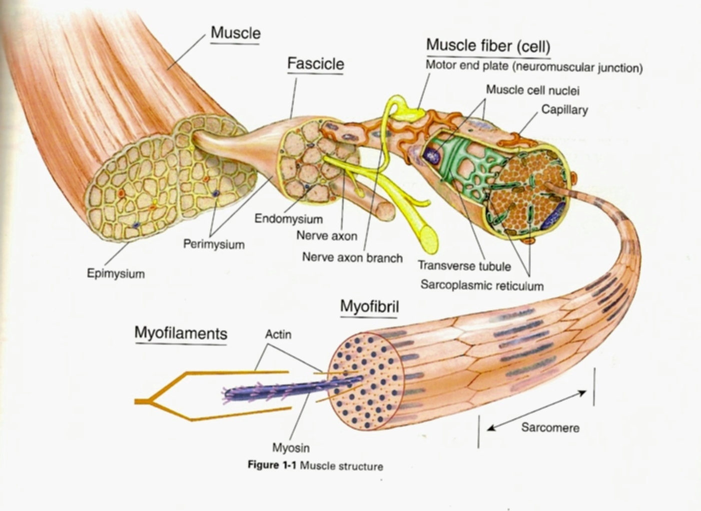
Structure of Skeletal Muscle
keletal muscle, a specialized contractile tissue found in animals, functions to move an organism’s body. It is composed of bundles of elongated muscle fibers, which are cylindrical and multi-nucleated. These fibers show a characteristic banding pattern with cross-striations of alternating light and dark bands.
Muscle Fibers and Cells
Each skeletal muscle fiber is formed from a chain of multinucleated muscle cells. These muscle cells are long and multinucleated. Each muscle cell has distinct regions when viewed under a microscope. These are known as sarcomeres, and give skeletal muscle a banded or striated appearance.
arcomeres
Each sarcomere is a complex of proteins, which operates to contract the muscle. Sarcomeres are formed from actin and myosin, as well as a number of associated helper proteins. The filaments seen between the dark bands are actin and myosin filaments. Actin is composed of many units of actin and takes the form of a twisting filament. Actin is accompanied by a number of proteins which help stabilize it and provide a pathway for muscle contraction. The two most important are tropomyosin and troponin. Tropomyosin surrounds the actin filament, and stops the heads of myosin from attaching. Troponin locks tropomyosin in place until receiving the signal to contract.
Connective Tissue Layers
Each skeletal muscle has three layers of connective tissue (called mysia) that enclose it, provide structure to the muscle, and compartmentalize the muscle fibers within the muscle. Each muscle is wrapped in a sheath of dense, irregular connective tissue called the epimysium. The epimysium also separates muscle from other tissues and organs in the area, allowing the muscle to move independently.
Fascicles
Inside each skeletal muscle, muscle fibers are organized into bundles, called fascicles, surrounded by a middle layer of connective tissue called the perimysium. This fascicular organization is common in muscles of the limbs; it allows the nervous system to trigger a specific movement of a muscle by activating a subset of muscle fibers within a fascicle of the muscle.
Endomysium
Inside each fascicle, each muscle fiber is encased in a thin connective tissue layer of collagen and reticular fibers called the endomysium. The endomysium surrounds the extracellular matrix of the cells and plays a role in transferring force produced by the muscle fibers to the tendons.
Tendons
At the ends of each skeletal muscle, a tendon connects the muscle to bone. This tendon connects directly to the epimysium, or collagenous outer covering of skeletal muscle. The tension created by contraction of the muscle fibers is then transferred though the connective tissue layers, to the tendon, and then to the periosteum to pull on the bone for movement of the skeleton.
Function of Skeletal Muscle
When you want to move your arm, your brain sends a nervous signal through your nerves. Each skeletal muscle receives the nervous impulse at neuromuscular junctions. These are places where nerves can stimulate an impulse in a muscle cell. The impulse travels down channels in the sarcolemma, the plasma membrane of skeletal muscle cells. At certain places in the membrane, there are channels that lead inside the cell. These carry the nervous impulse inside the cell..