Browse 3,344 knee anatomy stock photos and images available, or search for knee anatomy illustration or human knee anatomy to find more great stock photos and pictures.
Anatomy of the Knee Joint. The knee joint joins the thigh with the leg and consists of two articulations: one between the femur and tibia and one between the femur and patella. It is the largest joint in the human body. Illustration anatomy body. knee anatomy stock illustrations Anatomy of the Knee Joint.
Knee X-ray: A plain X-ray film of the knee is typically the best initial imaging test for most knee conditions. Magnetic resonance imaging (MRI scan): Using high-energy magnetic waves, an MRI scanner creates highly detailed images of the knee and leg.
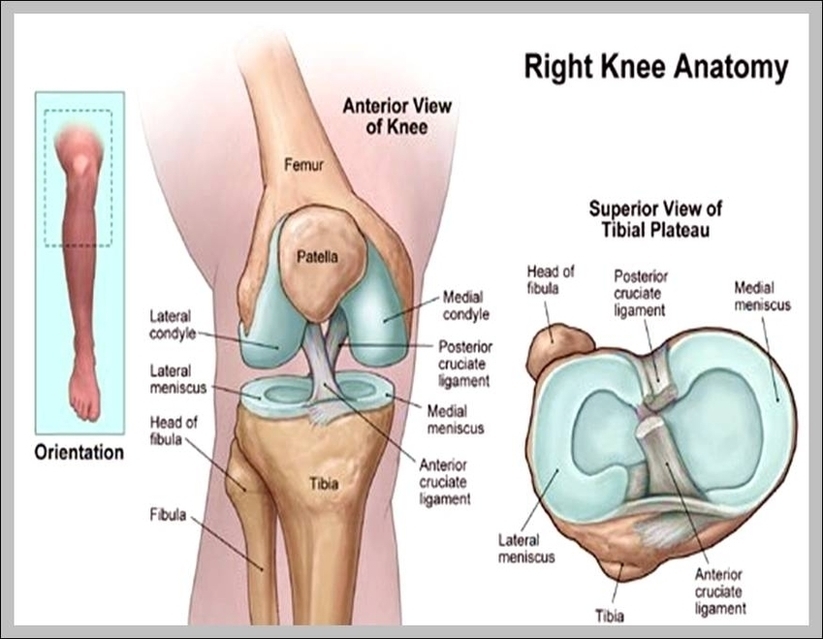
Pictures Of The Knee Anatomy Image
Posted inDiagrams
Pictures Of The Knee Anatomy Image
Post navigation
Previous Post
 Iliac Node Image
Next Post
Spine Structure Diagram Image