WebMD’s Knee Anatomy Page provides a detailed image and definition of the knee and its parts including ligaments, bones, and muscles. Skip to main content
X-rays of the knee are a commonly performed test to evaluate the knee joint. This is a front view of the knee joint, called an AP view of the knee. AP stands for anterior to posterior, meaning the image is directed from the front to the back of the knee joint.
X-rays of the knee are a commonly performed test to evaluate the knee joint. This is a front view of the knee joint, called an AP view of the knee. AP stands for anterior to posterior, meaning the image is directed from the front to the back of the knee joint.

Picture Of A Knee Cap Image
Posted inDiagrams
Picture Of A Knee Cap Image
Post navigation
Previous Post
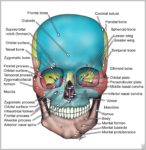 Skull Bone Anatomy Image
Next Post
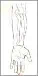
Supination Of Hand Image