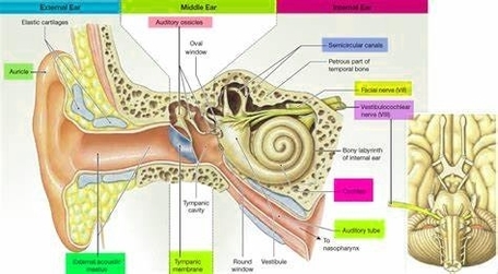
The inner ear, also known as the labyrinth, is the deepest part of your ear and plays a crucial role in hearing and maintaining balance. It consists of tiny bony structures filled with fluid. The inner ear is divided into three main parts: the cochlea, the semicircular canals, and the vestibule.
1. Cochlea
The cochlea is the auditory area of the inner ear that changes sound waves into nerve signals. It is made of a hollow bone shaped like a snail and divided into two chambers by a membrane. The chambers are full of fluid, which vibrates when sound comes in and causes the 30,000 tiny hairs lining the membrane to vibrate and send electrical impulses (sound signals) to the brain. The cochlea is about 9 millimeters wide at its widest point, and about 5 millimeters tall. If it could be uncoiled, the cochlea would be about 30 millimeters long.
2. Semicircular Canals
Also known as the labyrinthine, the semicircular canals rest on top of the cochlea, connected by the vestibule. There are three of them, and they line up at 90-degree angles to one another, which allows the brain to know which direction the head is moving. Like the cochlea, these canals are filled with fluid. They also contain small calcium crystals and tiny hairs that sense the movement of the fluid.
3. Vestibule
The vestibule is the central part of the bony labyrinth. It is separated from the middle ear by the oval window, and communicates anteriorly with the cochlea and posteriorly with the semicircular canals.
Inside the bony labyrinth lies the membranous labyrinth, which is also made up of three parts: the cochlear duct, the semicircular ducts, and the utricle and saccule.
4. Cochlear Duct
This triangle-shaped duct is located inside the bony labyrinth and creates two canals that sit above and below it. These two canalsthe scala vestibuli above the duct and the scala tympani below itare separated from the main duct by membranes.
5. Semicircular Ducts
This is where fluid, called endolymph, changes speed and direction when you move your head. Sensory receptors in these ducts detect this change and send information to your brain to help you maintain balance.
6. Utricle and Saccule
These are two small sacs located in the vestibule of the inner ear. They are filled with a jelly-like substance and tiny calcium carbonate crystals. When you move your head, these crystals move, causing the hairs in the utricle and saccule to bend. This sends a signal to your brain about the body’s position and movement.
The inner ear plays a key role in converting sound waves into electrical signals that the brain can interpret as sound, and in maintaining balance by sensing the position and movement of the head. Problems with the inner ear can result in hearing loss and balance issues..