A Chest Computed Tomography (CT) is a non-invasive diagnostic imaging procedure that uses special X-ray equipment to create detailed pictures, or scans, of areas inside the body. It is also sometimes referred to as a CAT scan (Computed Axial Tomography). It is used to visualize the heart, lungs, blood vessels, and bones in the chest region.
A normal chest CT scan reveals several key features:
1. Lungs and Airways: The lungs and airways appear normal in size and shape without any signs of inflammation. In a healthy lung, the diaphragm appears domed.
2. Pleura: The pleura, the thin membrane that lines the outer surface of the lungs and the inner surface of the chest wall, shows no signs of effusion (fluid) or thickening.
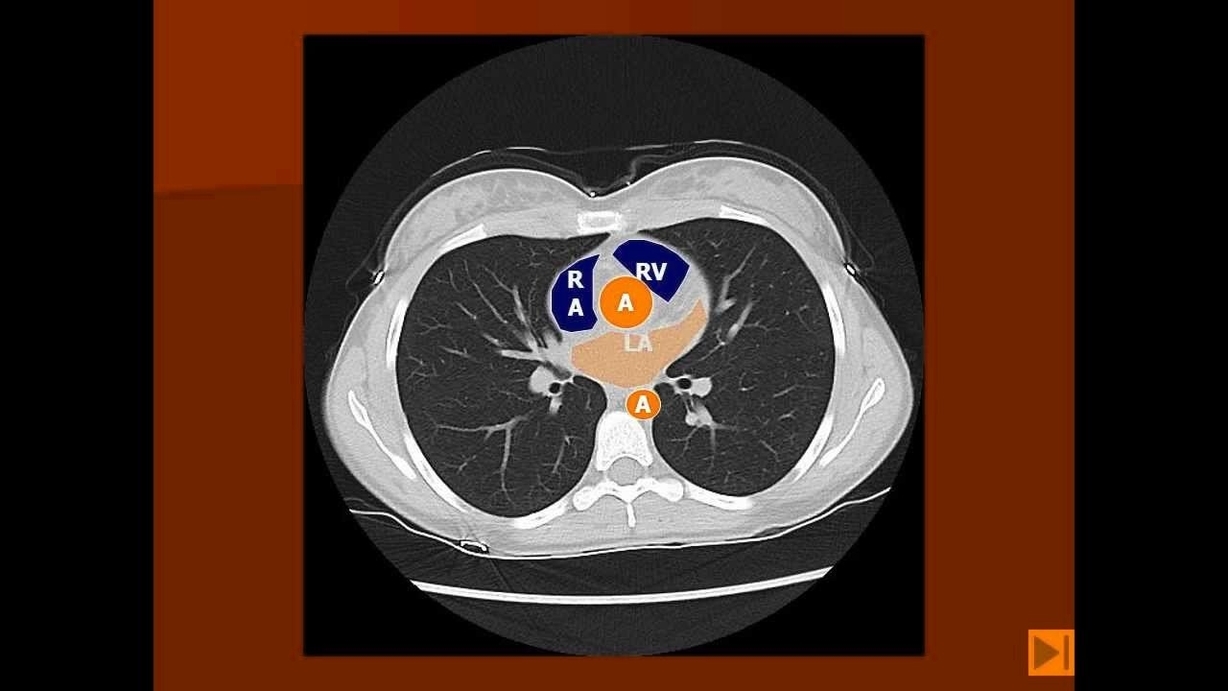
3. Heart: The heart size is normal without any signs of enlargement.
4. Pericardium: The pericardium, the double-walled sac containing the heart and the roots of the great vessels, shows no signs of effusion (fluid).
5. Mediastinum: The mediastinum, the central compartment of the thoracic cavity, and its structures have a normal configuration.
6. Chest Wall: The chest wall, which includes the ribs, muscles, and skin, is unremarkable.
A chest CT scan can be performed with or without contrast. Contrast is a special dye injected into the patient to make the structures inside the body more visible in the CT images. A normal CT chest with intravenous contrast is performed for a patient with sudden tearing central chest pain.
The rise of modern technology has allowed for the development of more advanced imaging techniques. For example, volumetric axial images can be obtained through the thorax without contrast media injection. These techniques provide a more detailed view of the chest’s anatomy, aiding in the diagnosis and treatment of various conditions.
In conclusion, a normal chest CT scan provides a detailed view of the chest’s anatomy, including the heart, lungs, blood vessels, and bones. It is a valuable tool in diagnosing and monitoring a wide range of health conditions. However, it is important to note that while a CT scan provides detailed images of the chest, it is only one piece of the puzzle. A comprehensive understanding of a patient’s health requires a combination of imaging results, physical examination, and medical history..