Each anatomical element was labeled on the 3 space planes: axial, frontal and sagittal. Anatomical structures of the face and oral cavity labeled on a MRI axial slice : pharynx, tong, tooth, masticator muscles… On the top menu, the user can select the labels to be displayed:
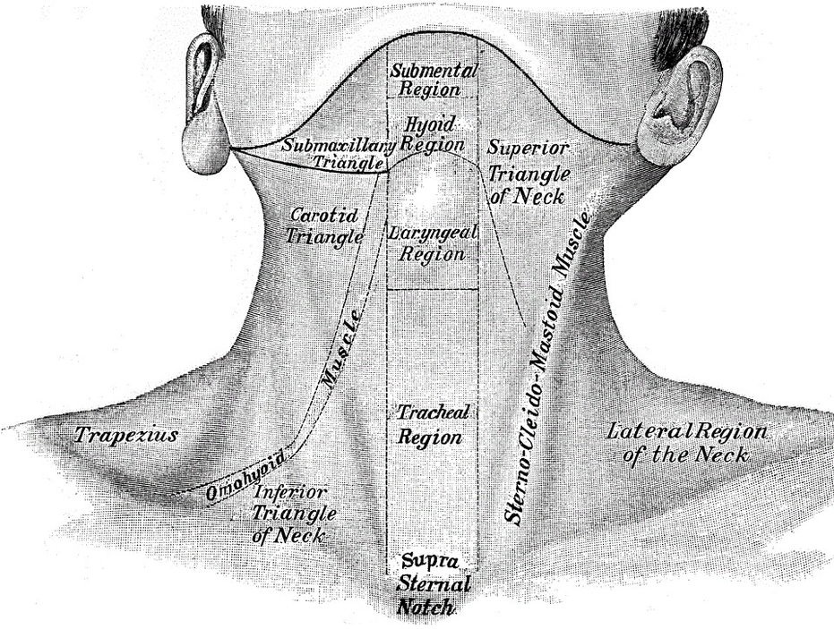
Bones of the Neck: The neck as you can see the neck is quite a complex structure. With seven cervical bones or vertebrae it has a great deal of ability for flexion, extension, and rotation. The bones that make up the neck are responsible for support and structure along with protection of the spinal cord as it exits the skull.
The bones of the face and neck were labeled using different colors to facilitate comprehension. The bone structures are rather more difficult to view on a weighted MRI T2 than on a CT-Scan: for more details on the bones of the face, please refer to the e-Anatomy module “Face-CT-Scan”.