The microscopic structure of bone is a fascinating topic that delves into the intricate details of how bones are formed and maintained.
Bone Tissue
Bone tissue, also known as osseous tissue, is unique in its hardness, a characteristic that is crucial for many of its functions. This hardness is due to the bone matrix, which consists of both organic and inorganic components. The organic component, known as osteoid, is laid down by bone cells called osteoblasts. The osteoid is then hardened with inorganic salts such as calcium and phosphate, a process known as mineralization.
Osteon: The Basic Microscopic Unit
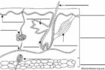
The basic microscopic unit of bone is an osteon, or Haversian system. Osteons are cylindrical structures that can measure several millimeters long and around 0.2 mm in diameter. Each osteon consists of lamellae of compact bone tissue that surround a central canal, the Haversian canal. This canal contains the bone’s blood supplies.
Compact Bone and Spongy Bone
The outer walls of the diaphysis, or shaft, of a long bone are composed of dense and hard compact bone. Compact bone is composed of a calcified bone matrix arranged in concentric rings. These rings contain cavities, or lacunae, filled with bone cells, or osteocytes. These osteocytes are interconnected by many minute passages, or canaliculi, which serve to distribute nutrients throughout the bone.
The wider section at each end of the bone, known as the epiphysis, is filled internally with spongy bone. Spongy bone, unlike compact bone, has a much less dense structure, which makes it lighter and more flexible.
Woven Bone and Lamellar Bone
Osteons can be arranged into woven bone or lamellar bone. Woven bone is characterized by the irregular organization of collagen fibers and is mechanically weak. It is found on the growing ends of an immature skeleton or, in adults, at the site of a healing fracture. Woven bone is replaced by lamellar bone during development.
Lamellar bone, in contrast, is highly organized in concentric sheets with a much lower proportion of osteocytes to surrounding tissue. The regular parallel alignment of collagen into sheets, or lamellae, causes lamellar bone to be mechanically strong. Lamellar bone makes up the compact or cortical bone in the skeleton, such as the long bones of the legs and arms.
Periosteum and Endosteum
On the outside of bones, there is a layer of cells that grow, repair, and remodel bone, which is part of the outer double-layered structure called the periosteum. The periosteum also contains blood vessels, nerves, and lymphatic vessels that nourish compact bone. Inside the bone, adjacent to the medullary cavity, is a layer of bone cells called the endosteum. These bone cells cause the bone to grow, repair, and remodel throughout life.
In conclusion, the microscopic structure of bone is a complex and dynamic system that allows for the growth, repair, and remodeling of bone throughout life. It is a testament to the remarkable adaptability and resilience of the human body..