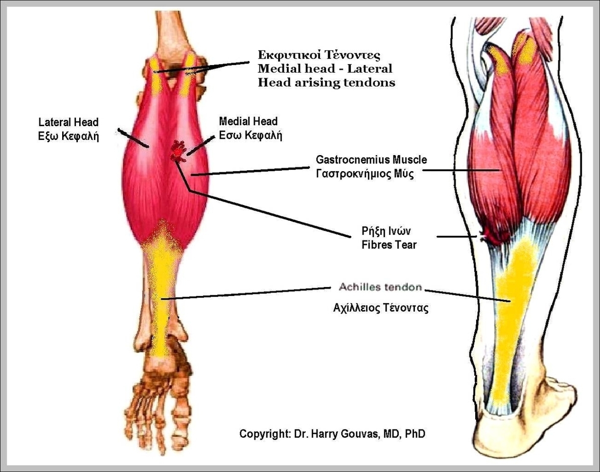
The gastrocnemius is located with the soleus in the posterior (back) compartment of the leg. The lateral head originates from the lateral condyle of the femur, while the medial head originates from the medial condyle of the femur .
Since the anterior compartment of the leg is lateral to the tibia, the bulge of muscle medial to the tibia on the anterior side is actually the posterior compartment. The soleus is superficial to the mid-shaft of the tibia. 10% to 30% of individuals have a sesamoid bone called the “fabella” in the lateral (outer) head of the gastrocnemius muscle.
Since the anterior compartment of the leg is lateral to the tibia, the bulge of muscle medial to the tibia on the anterior side is actually the posterior compartment. The soleus is superficial to the mid-shaft of the tibia. 10% to 30% of individuals have a sesamoid bone called the “fabella” in the lateral (outer) head of the gastrocnemius muscle.

Lateral Gastrocnemius Image
Posted inDiagrams

