The inner ear, the innermost part of the ear, plays a crucial role in hearing and balance. It consists of tiny bony structures filled with fluid. As sound waves travel from the outer to the inner ear, they create waves in the fluid of the inner ear, which in turn moves the tiny hairs in the ear that send sound or movement signals to the brain. Problems with this part of the ear can result in hearing loss and balance issues.
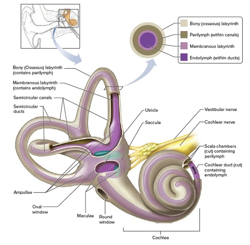
The inner ear is made up of the bony labyrinth and membranous labyrinth. The bony labyrinth comprises three components:
1. Cochlea: The cochlea is made of a hollow bone shaped like a snail and divided into two chambers by a membrane. The chambers are full of fluid, which vibrates when sound comes in and causes the 30,000 tiny hairs lining the membrane to vibrate and send electrical impulses (sound signals) to the brain. The cochlea is about 9 millimeters wide at its widest point, and about 5 millimeters tall. If it could be uncoiled, the cochlea would be about 30 millimeters long.
2. Semicircular Canals: Also known as the labyrinthine, the semicircular canals rest on top of the cochlea, connected by the vestibule. There are three of them, and they line up at 90-degree angles to one another, which allows the brain to know which direction the head is moving. Like the cochlea, these canals are filled with fluid. They also contain small calcium crystals and tiny hairs that sense the movement of the fluid.
3. Vestibule: The vestibule is the central part of the bony labyrinth. It is separated from the middle ear by the oval window, and communicates anteriorly with the cochlea and posteriorly with the semicircular canals.
Inside the bony labyrinth lies the membranous labyrinth, which is also made up of three parts:
1. Cochlear Duct: This triangle-shaped duct is located inside the bony labyrinth and creates two canals that sit above and below it. These two canalsthe scala vestibuli above the duct and the scala tympani below itare separated from the main duct by membranes. The membrane between the cochlear duct and the scala tympanialso known as the basilar membraneis where the primary hearing organ, the Organ of Corti, is located. The upper membrane is called Reissner’s membrane, which helps control the flow of fluid from the duct to the scala vestibuli.
2. Semicircular Ducts: This is where fluid, called endolymph, changes speed and direction when you move your head. Sensory receptors in these ducts detect this change and send information to your brain to help you maintain balance.
The inner ear is the last stop that sound waves make in a carefully orchestrated journey that starts from your outer ear. These waves travel from your outer ear through your middle ear to your inner ear. In the inner ear, the sound waves are converted into electrical energy, which your hearing nerve delivers to your brain as sound, making it possible for you to hear. At the same time, your inner ear monitors your movements, alerting your brain to changes so your brain can let your body know what to do to stay balanced..