
Human Skull Anatomy Study
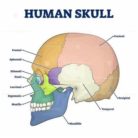
The human skull, a complex structure, serves as the body’s most vital protective mechanism, safeguarding the brain while supporting facial structures. It comprises 22 bones, or 29 when including the inner ear bones and hyoid bone. These bones are primarily connected by ossified joints known as sutures.
The skull is divided into two main parts: the neurocranium (braincase) and the viscerocranium (facial skeleton). The neurocranium encloses and protects the brain, with openings at the skull base for blood vessels and cranial nerves. The viscerocranium supports all facial structures.
Neurocranium
The neurocranium consists of the skullcap (calvarium) and the skull base. The calvarium is formed by the pairs of parietal bones and parts of the frontal bone as well as the occipital bone. The most significant sutures in the human skull are the coronal suture (between the frontal and parietal bone), the sagittal suture (dividing both the parietal bones), and the lambdoidal suture (running horizontally between the occipital bone and both parietal bones).
Viscerocranium
The viscerocranium comprises the facial bones that form the upper and lower jaws, nose, orbits, and other facial structures. It provides the bony support for the eyes, teeth, and structures of the face and provides openings for eating and breathing.
Cranial Fossae
The skull houses three cranial fossae: anterior, middle, and posterior. Each fossa contains different structures and has various openings for nerves and vessels.
1. Anterior Cranial Fossa: Contains the frontal lobe of the cerebral cortex, olfactory bulb, olfactory tract, optic nerve, and orbital gyri.
2. Middle Cranial Fossa: Houses the trochlear, abducens, oculomotor, ophthalmic, maxillary, mandibular nerves, pituitary gland, internal carotid artery, and temporal lobes of the brain.
3. Posterior Cranial Fossa: Contains the brainstem, facial, vestibulocochlear, glossopharyngeal, vagus, accessory, hypoglossal nerves, and internal jugular vein.
Facial Bones
The facial bones include the vomer, two inferior nasal conchae, two nasal, two maxillae, mandible, two palatine, two zygomatic, and two lacrimal bones. These bones form the nasal cavity, enclose the eyeballs, and support the teeth of the upper and lower jaws.
Conclusion
The study of the human skull anatomy is crucial in various fields such as medicine, anthropology, and forensic science. Understanding its complex structure and functions can provide insights into human evolution, health, and disease. It’s a fascinating area of study that continues to reveal the intricate design and functionality of the human body..